


Vol 28, No 1 (2025)
- Year: 2025
- Published: 06.02.2025
- Articles: 12
- URL: https://rjsvd.com/1560-9588/issue/view/9858
- DOI: https://doi.org/10.17816/dv.281

DERMATO-ONCOLOGY
Stages of training neural networks for classification and detection of skin neoplasms
Abstract
BACKGROUND: In recent years, neural networks have become an integral part of many fields, including medicine. However, the effectiveness of these models directly depends on the quality of the training data on which they are trained. Creating and maintaining a high-quality training dataset is a critical step in the development process of neural networks.
AIM: The aim of the research is to identify the key characteristics of the training database for the neural network that influence its subsequent sensitivity and specificity.
MATERIALS AND METHODS: A database of verified images of skin neoplasms was created to train a neural network to implement it in large-scale screening examinations. In the first phase of the study, a database was created to train a neural network to classify images of skin neoplasms (NSCa). Between 2017 and 2019, 7,680 digital images were collected from 6,892 patients with verified diagnoses: 5,316 (69,22%) confirmed by pathological examination, and 2,364 (30,78%)) confirmed clinically and dermatoscopically. A dataset containing 7,680 verified clinical images of skin neoplasms was created, and 1,680 images constituted the test sample for analyzing the model's effectiveness. The performance indicators of NSCa were as follows: sensitivity (Se): 70.47%; specificity (Sp): 79.86%; diagnostic accuracy (Ac): 74.68%. Due to the low sensitivity and specificity rates, the following steps were taken: (1) an additional round of training was conducted; (2) image quality control methods were developed; (3) a detection neural network was created, and (4) a new neural (NSCb) was established.
RESULTS: The neural network, trained on a verified dataset of clinical images of benign and malignant skin neoplasms and having undergone multiple rounds of training, operates with a sensitivity of 85.32–86.97% and a specificity of 87.59–88.92%. These rates exceed the sensitivity and specificity of skin neoplasm diagnoses made by non-oncological specialists using the naked eye, allowing for the use of this method in population screening. Following the retraining of the neural network and the establishment of NSCb, the creation of neural network, and the development of image quality control methods, an increase in the sensitivity and specificity of the neural network's performance was observed.
CONCLUSION: The use of artificial intelligence as a physician's assistant imposes quite high requirements on the performance parameters of the neural network. Mechanical learning, even on a large volume of verified data, did not achieve the desired results. The sequential work aimed at improving the parameters involved conducting an additional round of training, developing image quality control methods, and creating a detection neural network and a classification neural network. As a result, the trained neural network operates with a sensitivity of 85.32% to 86.97% and a specificity of 87.59% to 88.92%, which has enabled the use of the trained neural network as a tool for population screening.
 5-15
5-15


DERMATOLOGY
Folliculitis decalvans. Possibilities of combined therapy with 308 nm UVB: clinical cases
Abstract
Folliculitis decalvans is a rare neutrophilic cicatricial alopecia of the scalp. As it is a progressive inflammatory disease characterized by a gradual increase in the area of inflamed follicles with the formation of areas of cicatricial alopecia in the outcome. The most common clinical manifestations of folliculitis decalvans are follicular pustules, polytrichia, erosions and hemorrhagic crusts. At later stages dermal fibrosis predominates. The exact cause of folliculitis decalvans is not fully understood, but it is believed that Staphylococcus aureus may play a role in its development. Current knowledge regarding the pathogenesis of folliculitis decalvans suggests three stages including hyperactivation of innate immunity, bacterial infection and fibrosis as an outcome of the disease. The most frequently used treatments are topical steroids, topical antibiotics, and systemic antibacterial drugs, systemic isotretinoin, topical anti-inflammatory drugs, genetically engineered biological drugs, laser and surgical treatment, but all of the above methods usually result in only temporary remission. Phototherapy is an efficient therapy for a variety of skin diseases. We hypothesize that wavelengths in a range 308 nm eradicate bacteria more effectively than antibiotics in monotherapy. The combination of ultraviolet-B 308 nm, oral antibiotics, topical combined glucocorticoid drugs, topical and systemic application of bacteriophages, sensitives to the identified flora to the drugs have demonstrated an excellent tolerance and great outcomes in a series of cases.
16-26
Granuloma annulare: modern concepts of pathogenesis and pathogenetically based therapy (literature review)
Abstract
Granuloma annulare is a chronic, benign, non-infectious skin disorder that causes ring-shaped papules, most commonly on the hands and feet. The condition is considered rare, according to some data, it affects less than 0.04% of the population. However, in recent years there has been an increase in the incidence of dermatosis, which is associated with the high prevalence of infectious diseases, including infections caused by the SARS-CoV-2 coronavirus. The pathogenesis of granuloma annulare has not been fully deciphered; nevertheless, comparative analysis of a wide range of biomarkers in biopsies of affected skin has shown differences in the expression of genes involved in regulation of innate immunity, Th1 and Th2 lymphocytes, and Janus kinases. Associations of granuloma annulare with endocrine, immune, and autoimmune diseases, malignant neoplasms, as well as the triggering role of infections and vaccinations, are subjects of discussion, despite advances in understanding the molecular mechanisms of disease development.
The treatment of granuloma annulare presents quite complex challenge. Topical corticosteroids can be effective in localized forms of the disease. Calcineurin inhibitors can be used in combination with them. For disseminated forms of the dermatosis, narrowband medium wave ultraviolet therapy at 311 nm, PUVA, and 308 nm excimer ultraviolet light therapy are prescribed. There are descriptions of the effectiveness of antibacterial drugs.
Currently, TNF-α inhibitors, JAK inhibitors and IL-23 inhibitors are proposed as targeted therapy for granuloma annulare. The basis for this recommendation is the latest information about the pathogenesis of granuloma annulare.
The presented literature review systematizes modern concepts of molecular and cellular mechanisms of granuloma annulare development, infectious and non-infectious triggers of inflammation, the role of endocrine, immune and oncological pathology as predisposing factors. Significant importance is given to the analysis of treatment methods of the disease.
27-40
Differential diagnosis of ulcerative lesions in a patient with pyoderma gangrenosum in association with ulcerative colitis and multiple endocrine neoplasia
Abstract
Differential diagnosis of chronic ulcers of non-infectious nature remains a complex interdisciplinary problem.
Pyoderma gangrenosum is a rare autoinflammatory disease, which is referred to the group of neutrophilic dermatoses and is clinically manifested by the rapid development of painful, long-term persistent, recurrent skin ulcers with irregular, undermined borders of red-violet colour and surrounding erythema. Annually from 3 to 10 cases of pyoderma gangrenosum per million people are reported. To the present time, the etiology and pathogenesis of pyoderma gangrenosum remain completely unstudied, and the diagnosis is complicated by the rare incidence of the disease and non-specificity of cutaneous manifestations.
In this article the observation of a long-term course of pyoderma gangrenosum in a 58-year-old female with ulcerative colitis and multiple endocrine neoplasia type I. The differential diagnosis between pyoderma gangrenosum with necrotising ulcerative vasculitis was performed. Based on the results of the comprehensive examination, the final diagnosis of pyoderma gangrenosum was established.
This clinical observation demonstrates the necessity for the detailed examination of patients in order to make a correct diagnosis and prescribe adequate treatment on time.
41-52
Laser therapies for the treatment of necrobiosis lipoidica: literature review and own experience
Abstract
Necrobiosis lipoidica is a rare, chronic skin disease of vascular-exchange character from the group of localised skin lipoidosis, characterised by collagen degeneration. The main methods of treatment of this dermatosis do not always have a positive effect and do not lead to long-term remission. In this regard, in recent years, new methods of the treatment for necrobiosis lipoidica have appeared, in particular laser treatment. The aim of this study is to investigate the efficacy and safety of laser therapy for necrobiosis lipoidica, as well as to report on our own experience of using IPL therapy for necrobiosis lipoidica.
A review of scientific publications on the topic over the past 5 years on platforms such as PubMed and Google Scholar was conducted. Laser treatments used in the selected studies included CO2 laser, dye laser and broadband intense pulsed light radiation (IPL therapy). Publications that used combined treatments were excluded.
The description of our own experience of using intense pulsed light for the treatment of necrobiosis lipoidica in four patients with a positive effect in the form of reduction of telangiectasias, discoloration and flattening of the peripheral boundaries is presented. In connection with the increasing interest to laser methods of therapy of various dermatoses, we consider it important to study and apply them in the treatment of necrobiosis lipoidica, which may help to develop a new approach to the treatment of necrobiosis lipoidica.
53-62
Development and application of a multicomponent film biocoating based on sodium carboxymethylcellulose to study the antibacterial activity of microflora in the oral fluid of patients with pemphigus vulgaris in vitro
Abstract
BACKGROUND: A special place among dermatological diseases is occupied by dermatoses affecting the oral mucosa, in particular pemphigus vulgaris. Constant damage to the mucous membrane, combined with the presence of abundant microflora in the oral cavity, leads to rapid variability in the primary or pathognomonic manifestations of specific diseases, making them look similar. Erosion and ulcers in the oral cavity are very difficult to treat and are accompanied by severe pain. The role of the oral microflora in the development and progression of pemphigus vulgaris has not been fully studied, however, it is known that the microflora of patients differs significantly from that of healthy individuals. Today, much attention is paid to studying the composition, properties and role of the microflora of the oral mucosa in the manifestation and course of pemphigus vulgaris.
AIM: determination of the most effective combination of a three-component film biocoating based on sodium carboxymethylcellulose for patients with pemphigus vulgaris, by assessing and comparatively in vitro comparison of quantitative and qualitative characteristics of the oral microflora, as well as local oral protective factors (lysozyme titer, phagocytosis index neutrophils and the level of the secretory fraction of immunoglobulin class A)
MATERIALS AND METHODS: A cross-sectional single-center study was performed among patients with pemphigus vulgaris (n=12). Oral fluid was collected by washing (rinsing) 4.5 ml of physiological solution from the oral mucosa. Subsequently, in the laboratory, a series of serial dilutions were prepared, some of which were inoculated by quantitative sectoral sowing on media intended for the cultivation of aerobic and anaerobic microbes. Microbiological and immunological sensitivity to films with different concentrations of mometasone furoate (20–40–80 mg) and propolis (2.5–5–7.5–10%) was determined.
RESULTS: The most pronounced antibacterial activity was observed in the following 2-component forms (gel film and propolis at a concentration of 7.5%; gel film and mometasone furoate in an amount of 20 mg) and in 3-component gel films (gel film, propolis at a concentration of 5% and mometasone furoate 20 mg). According to the results of a comparative quantitative assessment of local factors protecting the oral cavity in patients with pemphigus vulgaris, their positive dynamics, characterized by the restoration of immunodeficiency, were most influenced by the three-component composition, including a gel film, propolis 5% and mometasone furoate 80 mg.
CONCLUSION: The three-component composition of the film biocoating based on carboxymethylcellulose significantly reduces the quantitative and improves the qualitative indicators of microorganisms in the oral cavity, thereby having a positive effect on the treatment process for patients with pemphigus vulgaris.
63-74
Multisystem Langerhans cell histiocytosis in adults: the significance of cutaneous manifestations in early diagnosis
Abstract
Langerhans cell histiocytosis is a rare disease characterized by the proliferation of Langerhans cells in various organs and tissues, including the skin, bones, lungs, and pituitary gland. The precursor cell, as recent studies have shown, is the myeloid dendritic cell. The severity of the disease is mainly determined by the extent and nature of organ involvement, as well as the number of systems affected. Diagnosis of Langerhans cell histiocytosis requires a comprehensive approach (clinical evaluation, histological analysis, immunohistochemical studies, radiological methods and molecular genetic tests).
This article presents a clinical case of a 28-year-old female patient with multisystem Langerhans cell histiocytosis, whose nonspecific skin eruptions were misdiagnosed as seborrheic dermatitis for five years. An inadequate reaction to glucocorticosteroid treatment, along with the presence of comorbid conditions such as diabetes insipidus and severe bullous lung lesions, necessitated additional diagnostic investigations. The final diagnosis was established after immunohistochemical examination of skin and lung biopsies, which revealed the expression of CD68 and CD1a in infiltrating cells.
It is important to emphasize that skin manifestations are often the first symptom of a multisystem process. Early recognition of Langerhans cell histiocytosis and timely diagnosis can significantly affect the course of the disease and improve the patient's quality of life.
75-86
The effect of phototherapy on the expression of innate immunity genes in patients with psoriasis
Abstract
BACKGROUND: Phototherapy is one of the most effective methods in the treatment of psoriasis, but the mechanism of its action on innate immunity has not been studied.
AIM: Investigation of the local expression profile of innate immunity factors in patients with psoriasis during phototherapy.
MATERIALS AND METHODS: The study included 31 patients diagnosed with inpatient psoriasis vulgaris. The material for the study was obtained from areas of affected and unaffected skin. Patients with vulgar psoriasis received a course of UVB-311 nm phototherapy lasting from 5 to 7 weeks with a total dose of 35.2 to 44.6 J/cm2. There were 30 healthy people in the control group. Gene expression analysis was performed before treatment and at the end of the phototherapy course. The data obtained were statistically processed.
RESULTS: According to the results of the study, gene expression data were obtained: for example, increased expression of the TLR2 and TLR9 genes was observed in the main group after treatment, as well as in samples of unaffected skin from patients. The increased level of the TLR4 gene expression was registered in unaffected skin samples from patients with psoriasis. The expression of the β-defensin 1 gene was elevated in unaffected skin and post-treatment skin. For the cathelicidin gene, there is a difference between the groups of affected and unaffected skin samples before treatment. The expression level of the IL-13 gene was higher before treatment.
CONCLUSION: The revealed local imbalance of factors of innate immunity can lead to a more severe course of the disease. The course of phototherapy leads to normalization of the expression profile of receptor and effector molecules of innate immunity, which leads to a stable positive clinical effect.
87-94
Determination of melanin levels in patients with vitiligo
Abstract
BACKGROUND: Vitiligo is an urgent problem for both patients and the scientific community of dermatologists. In this regard, there are many studies aimed at finding new methods of therapy for this disease, providing repigmentation of foci and stabilization of the process, as well as available methods of treatment control to objectify the results in dynamics.
AIM: To assess the melanin level in hypopigmentation foci in vitiligo patients before and during treatment using mexametry, as well as on the surrounding healthy skin, to study the possibility of using this method as a control of response to therapy.
MATERIALS AND METHODS: 17 vitiligo patients (10 female, 7 males, mean age 37.9±2.9, disease duration 16.5±2.3 years) participated in the dynamic follow-up. Patients were measured melanin level in depigmentation foci and on the surrounding healthy skin in 19 localizations by mexametry before treatment and after 3 months of therapy. The study was carried out on the Soft plus device. The degree of repigmentation was also assessed clinically using the visual analog scale of repigmentation (G0–G4).
RESULTS: According to mexametry data, the most significant differences between the melanin level in vitiligo foci and on the surrounding healthy skin of the upper and lower extremities, on the skin of the trunk (p <0.001). In 3 months after therapy a significant increase of melanin in vitiligo foci according to mexametry data was observed in the elbows ― from 2 u to 13 u, shoulders ― from 2 u to 17 u and knees ― from 5 u to 18 u. Increased melanin levels according to mexametry correlated with the clinical picture. Thus, these areas showed a G4 excellent (repigmentation over 75%) and G3 very good response (repigmentation 50–75%) on the visual analog scale of repigmentation. There were no correlations between skin melanin levels and age, sex.
CONCLUSION: In the course of treatment there was an increase in melanin level in vitiligo foci according to mexametry, which correlated with the clinical picture, and testified to the effectiveness of therapy. On the basis of the obtained results we can conclude that mexametry is a good objective method of monitoring the effectiveness of therapy in patients with vitiligo in dynamics.
95-102
COSMETOLOGY
The use of preparations based on hyaluronic acid modified with amino acids and preparations based on micronized collagen in injection cosmetology
Abstract
Skin condition often affects a person's emotional state, social interaction, and quality of life in general. Inevitable skin ageing, which is a long-term multifactorial process, includes transformation of tissue and cellular homeostasis, impaired proteostasis, decreased immunity, impaired DNA repair and other pathological processes. Correction of age-related changes in facial skin currently remains one of the most urgent tasks of modern aesthetic medicine.
Aesthetic medicine is one of the most dynamically developing areas of modern healthcare. Today, injectable cosmetology provides an opportunity for a pathogenetic approach to correcting age-related skin changes and solving a number of aesthetic problems. Most often, preparations containing hyaluronic acid, micronized collagen, vitamins, amino acids, and trace elements are used for this purpose.
This article presents an analysis of literature data devoted to the study of modern aspects of the use and effectiveness of injectable preparations based on hyaluronic acid modified with amino acids and preparations based on micronized collagen in aesthetic cosmetology.
103-113
CHRONICLES
Chronicles of A.I. Pospelov Moscow Society of Dermatovenerologists and Cosmetologists (MSDС was founded on October 4, 1891) Bulletin of the MSDС № 1159
Abstract
On October 15, 2024, the regular, 1159th meeting of the Moscow Society of Dermatovenerologists and Cosmetologists named after A.I. Pospelov was held.
The meeting was held in person, in which 110 people took part. 67 applications were submitted for membership in the MODV, 61 of which were from residents and employees of the V.A. Rakhmanov Clinic of Skin and Venereal Diseases.
The clinical part of the meeting presented two reports, the topics of which concerned skin sarcoidosis (clinical cases) and successful therapy of suppurative hidradenitis with secukinumab. The topics of the reports of the scientific part of the meeting were devoted to the expanded capabilities of broadband IPL light in dermatology, the evolution of views on comedogenesis in acne, as well as the real results of psoriasis therapy with netakimab. Suppurative hidradenitis is a chronic autoinflammatory skin disease, in the development of which a hereditary predisposition plays a leading role. Treatment depends on the severity of the process - from local therapy at the initial stage of the disease to the prescription of systemic antibiotics, drugs of genetic engineering biological therapy for moderate severity and surgical removal of the affected area with subsequent plastic surgery in severe cases. The topic of suppurative hidradenitis was also touched upon in the scientific part of the meeting - when discussing broadband IPL light, in particular, the effectiveness of its use in localizing the process in the armpit and groin areas. Other topics of scientific reports covered not only the problems of comedogenesis in acne, but also expanded the understanding of the role of drugs of genetic engineering biological therapy.
114-120
PHOTO GALLERY
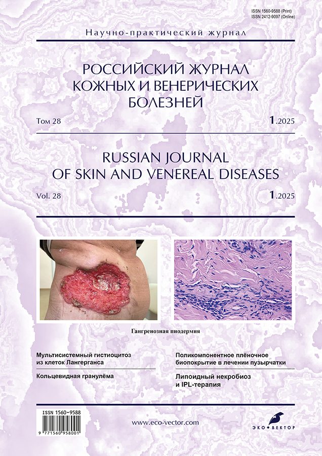
Photogallery. Pyoderma gangrenosum
Abstract
Pyoderma gangrenosum is a rare autoinflammatory disease belonging to the group of neutrophilic dermatoses. It is characterized by the formation of painful, rapidly progressing ulcerative necrotic lesions with covered edges of a bluish hue and pronounced surrounding erythema. Despite its historical name, pyoderma gangrenosum is not gangrene or pyoderma, which highlights the complexity of its pathogenesis and clinical course.
The pathophysiology of the disease includes dysregulation of innate immunity, genetic mutations, and neutrophil dysfunction.
Pyoderma gangrenosum often develops against the background of somatic diseases. In more than half of patients, it is associated with inflammatory bowel diseases (Crohn's disease, ulcerative colitis), hematological disorders (myeloma, chronic lymphocytic leukemia), as well as rheumatological diseases (rheumatoid arthritis). This fact makes pyoderma gangrenosum an important interdisciplinary disease that requires an integrated approach to diagnosis and treatment.
Ulcerative lesions are mainly localized on the lower extremities, however, their occurrence is also possible on the trunk, upper extremities, face and scalp, which significantly complicates the diagnosis and differentiation with other skin diseases.
Diagnosis of the disease is based on clinical manifestations, exclusion of other causes of ulcerative necrotic lesions (infectious, vascular and other processes), as well as histological examination data revealing abundant neutrophil infiltration of the dermis and hypodermis. The treatment of pyoderma gangrenosum includes suppressive drugs and targeted therapy using genetically engineered biological drugs (GIBPS) aimed at controlling the inflammatory process. The approach to therapy is determined individually, depending on the severity of the course and the presence of concomitant diseases.
We present the variability of clinical manifestations of pyoderma gangrenosum in order to improve timely diagnosis and adequate treatment (from the archive of The First Sechenov Moscow State Medical University, the photographs are published for the first time with the permission of the institution's administration).
121-129
Регистрационный номер и дата принятия решения о регистрации СМИ: серия ПИ № ФС 77 - 86501 от 11.12.2023 г.
СМИ зарегистрировано Федеральной службой по надзору в сфере связи, информационных технологий и массовых коммуникаций (Роскомнадзор).
Регистрационный номер и дата принятия решения о регистрации СМИ: серия ЭЛ № ФС 77 - 80653 от 15.03.2021 г.