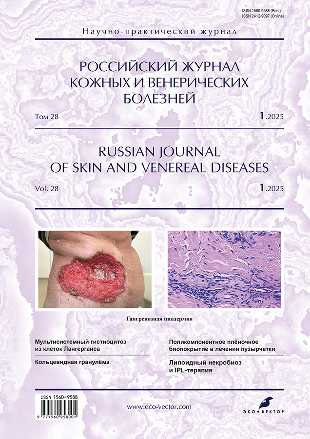
Photogallery. Pyoderma gangrenosum

封面


如何引用文章

全文:

开放存取 开放存取
受限制的访问 ##reader.subscriptionAccessGranted##
受限制的访问 订阅或者付费存取

详细

Pyoderma gangrenosum is a rare autoinflammatory disease belonging to the group of neutrophilic dermatoses. It is characterized by the formation of painful, rapidly progressing ulcerative necrotic lesions with covered edges of a bluish hue and pronounced surrounding erythema. Despite its historical name, pyoderma gangrenosum is not gangrene or pyoderma, which highlights the complexity of its pathogenesis and clinical course.

The pathophysiology of the disease includes dysregulation of innate immunity, genetic mutations, and neutrophil dysfunction.

Pyoderma gangrenosum often develops against the background of somatic diseases. In more than half of patients, it is associated with inflammatory bowel diseases (Crohn's disease, ulcerative colitis), hematological disorders (myeloma, chronic lymphocytic leukemia), as well as rheumatological diseases (rheumatoid arthritis). This fact makes pyoderma gangrenosum an important interdisciplinary disease that requires an integrated approach to diagnosis and treatment.

Ulcerative lesions are mainly localized on the lower extremities, however, their occurrence is also possible on the trunk, upper extremities, face and scalp, which significantly complicates the diagnosis and differentiation with other skin diseases.

Diagnosis of the disease is based on clinical manifestations, exclusion of other causes of ulcerative necrotic lesions (infectious, vascular and other processes), as well as histological examination data revealing abundant neutrophil infiltration of the dermis and hypodermis. The treatment of pyoderma gangrenosum includes suppressive drugs and targeted therapy using genetically engineered biological drugs (GIBPS) aimed at controlling the inflammatory process. The approach to therapy is determined individually, depending on the severity of the course and the presence of concomitant diseases.

We present the variability of clinical manifestations of pyoderma gangrenosum in order to improve timely diagnosis and adequate treatment (from the archive of The First Sechenov Moscow State Medical University, the photographs are published for the first time with the permission of the institution's administration).

全文:

受限制的访问

作者简介

Olga Grabovskaya

The First Sechenov Moscow State Medical University

编辑信件的主要联系方式.
Email: rjdv@eco-vector.com
ORCID iD: 0000-0002-5259-7481
俄罗斯联邦, Moscow

Diana Kusraeva

The First Sechenov Moscow State Medical University

Email: rjdv@eco-vector.com
ORCID iD: 0000-0002-5633-7986
俄罗斯联邦, Moscow

Anna Bobkova

The First Sechenov Moscow State Medical University

Email: rjdv@eco-vector.com
ORCID iD: 0000-0003-3611-0917
俄罗斯联邦, Moscow

参考

补充文件

附件文件
动作
1. JATS XML
2. Fig. 1. Patient B., 51 years old, ulcerative (classic) pyoderma gangrenosum, associated with rheumatoid arthritis: a ― ulcer on the back measuring 28×56 cm with sharply circumscribed, undermined, painful borders and perilesional erythema; the bottom of the ulcer is covered with hemorrhagic purulent exudate, occasionally with fibrin granulations; b ― healed pyoderma gangrenosum, an extensive hypertrophic scar of irregular shape with uneven edges in the chest region is presented; c ― multiple atrophic scars on the site of healed ulcers; deep ulcer on the posterior surface of the shin measuring 5×8 cm; the calcaneal tendon is visualized on the bottom of the ulcer.

下载 (1MB)
3. Fig. 2. Patient M., 59 years old, ulcerative (classic) pyoderma gangrenosum, associated with a tumor of the ascending colon: a ― multiple ulcers (n=5) of the abdomen up to 2.5 cm in size with undermined bluish color borders and perilesional erythema; the bottom of the ulcers is covered with scarce fibrin granulations; b ― ulcerative defect on the scrotum measuring 3×5 cm, of irregular shape, the bottom of the ulcer is covered with purulent exudate.

下载 (865KB)
4. Fig. 3. Patient C., 53 years old. Ulcerative (classic) pyoderma gangrenosum: ulcerative defect of the lateral surface of the shin with a transition to the ankle joint area, with clear contours, roller-like raised edges of a purplish-cyanotic color; the bottom of the ulcer is covered with hemorrhagic―purulent exudate, in places with fibrin granulations, and a zone of pink erythema around the ulcer (a). Vegetative (superficial granulomatous) pyoderma gangrenosum: extensive ulcer of the lateral surface of the trunk with a transition to the left buttock, with undermined, irregular, erythematous-violaceous edges and perilesional erythema; the bottom of the ulcer is covered with hemorrhagic purulent exudate (b).

下载 (1MB)
5. Fig. 4. Patient V., 89 years old, ulcerative (classic) pyoderma gangrenosum, associated with multiple myeloma: a ― inner surface of the thigh ulcer with clear contours, undermined, painful borders and perilesional erythema; the bottom of the ulcer is covered with purulent hemorrhagic exudate, occasionally with fibrin granulations; b ― multiple ulcers with erythematous border on the shin measuring up to 3 cm in diameter, of circular shape, with undermined, painful borders and perilesional erythema; c ― painful pustules in the chest area (early pustular pyoderma gangrenosum).

下载 (1MB)
6. Fig. 5. Patient L., 69 years old, ulcerative (classic) pyoderma gangrenosum: a ― multiple ulcers with overhanging edge; the bottom of the ulcers is covered with purulent exudate, scarce purulent crusts; b ― gangrenous pyoderma rashes at different stages of development of the elements ― violet papule with central vesiculation (1), ulcers with undermined borders up to 1.5 cm in diameter (2); c ― ulcers on the forearm area of with undermined borders are presented.

下载 (1MB)
7. Fig. 6. Patient K., 61 years old, ulcerative (classic) pyoderma gangrenosum, with signs of artificial dermatitis in a patient with rheumatoid arthritis: multiple superficial ulcers up to 12–15 cm in size, prone to fusion with the formation of a vast irregular lesion with sharply circumscribed, undermined, painful borders and perilesional erythema; the bottom of the ulcers is covered with purulent exudate, occasional fibrin granulations, and a white plaque (microbiological culture analysis ― Candida+), the bottom of the ulcer is covered with purulent exudate.

下载 (621KB)
8. Fig. 7. Patient N., 63 years old, ulcerative (classic) pyoderma gangrenosum: the ulcer located on the lateral surface of the abdomen measuring 23×45 cm, with sharply circumscribed, undermined, painful borders and perilesional erythema; the bottom of the ulcers is covered with purulent exudate.

下载 (469KB)
9. Fig. 8. Patient A., 70 years old, ulcerative (classic) pyoderma gangrenosum: lateral malleolar ulcer, with sharply circumscribed, undermined, painful borders and perilesional erythema; the bottom of the ulcer is covered with hemorrhagic purulent exudate, occasionally with fibrin granulations.

下载 (633KB)
10. Fig. 9. Patient R., 64 years old, ulcerative (classic) pyoderma gangrenosum against the background of Rituximab for B-cell chronic lymphocytic leukemia: ulcer with erythematous border on the shin about 4×7 cm, with undermined, painful borders; the bottom of the ulcer is covered with hemorrhagic purulent crust.

下载 (449KB)
11. Fig. 10. Patient N., 59 years old, vegetative (superficial granulomatous) pyoderma gangrenosum: necrotic lesion about 12×13 cm on the lateral surface of the trunk, with purple undermined and rugged borders; the bottom of the ulcer is covered with hemorrhagic purulent exudate, occasionally with fibrin granulations.

下载 (458KB)
12. Fig. 11. Patient N., 45 years old, ulcerative (classic) pyoderma gangrenosum, edema and peri-ulcer scarring: undermined borders and perilesional erythema are no longer present, fat and fascia are visible at the base of the ulcers.

下载 (589KB)
13. Fig. 12. Patient D., 48 years old, vegetative (superficial granulomatous) pyoderma: ulcer on the shin, irregularly shaped, measuring 21×32 cm, sharply delimited from the surrounding apparently healthy skin and raised above its surface and perilesional erythema, the bottom of the ulcer is covered with hemorrhagic purulent exudate.

下载 (568KB)
14. Fig. 13. Patient C., 61 years old, ulcerative pyoderma gangrenosum: an irregular circular shin ulcer, with sharply circumscribed, undermined, painful borders; the bottom of the ulcer is covered with hemorrhagic purulent exudate, occasionally with fibrin granulations.

下载 (622KB)

版权所有 © Eco-Vector, 2025

许可 URL: https://eco-vector.com/for_authors.php#07

СМИ зарегистрировано Федеральной службой по надзору в сфере связи, информационных технологий и массовых коммуникаций (Роскомнадзор).
Регистрационный номер и дата принятия решения о регистрации СМИ: серия ПИ № ФС 77 - 86501 от 11.12.2023 г
СМИ зарегистрировано Федеральной службой по надзору в сфере связи, информационных технологий и массовых коммуникаций (Роскомнадзор).
Регистрационный номер и дата принятия решения о регистрации СМИ: серия ЭЛ № ФС 77 - 80653 от 15.03.2021 г
.