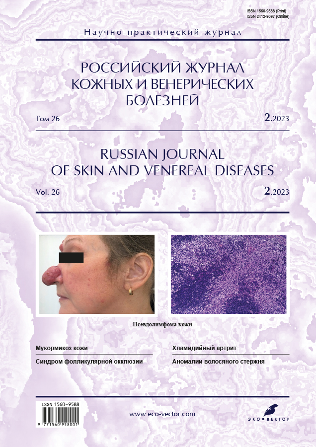
Photo gallery. Rare dermatoses. Part I
- Authors: Snarskaya E.S.1, Teplyuk N.P.1, Semiklet J.M.1
-
Affiliations:
- I.M. Sechenov First Moscow State Medical University (Sechenov University)
- Issue: Vol 26, No 2 (2023)
- Pages: 213-219
- Section: PHOTO GALLERY
- Submitted: 20.03.2023
- Accepted: 30.03.2023
- Published: 21.05.2023
- URL: https://rjsvd.com/1560-9588/article/view/321499
- DOI: https://doi.org/10.17816/dv321499
- ID: 321499
Cite item
Abstract
Orphan dermatoses are caused by both genetic causes and a number of autoimmune, inflammatory and infectious processes in the body. Congenital giant nevus is associated with serious complications: malignant melanoma, CNS lesions (neurocutaneous melanosis). Multiple epidermoid cysts are often a component of Gardner's syndrome. Angiolymphoid hyperplasia with eosinophilia, a vascular tumor formation with proliferation of histiocytoid endothelial cells, with severe lymphocytic and eosinophilic infiltration, recurs in 1/3 of cases. Morbigan's disease is persistent erythema and swelling of the middle third and upper part of the face. Skin mastocytosis in children is prone to spontaneous resolution during puberty. Squamous cell skin cancer accounts for 20% of all skin cancers and has a high risk of recurrence, metastasis, and death. Buschke's scleroderma is a scleroderma-like disease that causes progressive fibrous-mucinous thickening of the skin affecting the neck, shoulders, proximal upper extremities, and face. Fulminant (lightning) acne is a sharply exacerbated form of acne vulgaris, characterized by an acute onset and the appearance of confluent abscesses. Cases of acne fulminans have been observed during treatment with isotretinoin. Norwegian (crustose) scabies is associated with a compromised immune response, which allows the mites to multiply and can number in the millions. Pretibial myxedema (thyroid dermopathy) is a rare extrathyroid manifestation of Graves' disease along with thyrotoxicosis, thyroid hypertrophy, ophthalmopathy, and acropachy. Porokeratosis Mibelli predisposing factors of development are immunosuppressive conditions, malignancy is possible. Osteomas are formations from bone tissue in the dermis or subcutaneous fat as a result of metaplasia of fibroblasts into osteoblasts, or differentiation of primitive mesenchymal cells into osteoblasts and their migration to an ectopic site. Sarcoidosis of the skin is characterized by a variety of clinical forms, rarely observed before 40 years. Xanthomas with histiocytosis X ― a violation of lipid metabolism. The rural type of leishmaniasis (acute necrotizing) has a short incubation period, a significant spread of rashes and healing with the formation of scar tissue. The giant condyloma of Buschke–Levenshtein is characterized by progressive growth, large size and recurrent course, it can transform into squamous cell carcinoma. Degos disease ― malignant atrophic papulosis ― a rare disease caused by blockage of the arteries and veins.
Keywords
Full Text
Fig. 1. Patient A., 3 months. Congenital giant nevus of the skin of the foot.
Fig. 2. Patient A., 61 years. Multiple epidermal cysts of the scrotum.
Fig. 3. Patient A., 27 years. Angiolymphoid hyperplasia with eosinophilia in the ear area.
Fig. 4. Patient B., 30 years. Morbigan's Disease.
Fig. 5. Patient D., 1 year. Mastocytomas.
Fig. 6. Patient K., 57 years. Squamous cell skin cancer of the vulva.
Fig. 7. Patient K., 50 years. Adult Buschke scleredema.
Fig. 8. Patient R., 11 years. Simple contact dermatitis (contact with jellyfish).
Fig. 9. Patient M., 28 years. Complication after fulminant acne.
Fig. 10. Patient В., 54 years. Norwegian scabies.
Fig. 11. Patient E., 56 years. Pretibial myxedema.
Fig. 12. Patient К., 15 years. Porokeratosis Mibelli: a ― lower extremities; b ― image fragment.
Fig. 13. Patient M., 35 years. Multiple osteomas of the forehead skin.
Fig. 14. Patient M., 26 years. Sarcoidosis of the forehead skin.
Fig. 15. Patient M., 60 years. Xanthomas in histiocytosis X.
Fig. 16. Patient Н., 35 years. Rural type of leishmaniasis.
Fig. 17. Patient О., 47 years. Giant Buschke–Levenstein condyloma, multiple scrotal papillomas.
Fig. 18. Patient С., 35 years. Degos disease.
About the authors
Elena S. Snarskaya
I.M. Sechenov First Moscow State Medical University (Sechenov University)
Email: snarskaya-dok@mail.ru
ORCID iD: 0000-0002-7968-7663
SPIN-code: 3785-7859
MD, Dr. Sci. (Med.), Professor
Russian Federation, 119435, Moscow, st. Bolshaya Pirogovskaya, 4/1Natalya P. Teplyuk
I.M. Sechenov First Moscow State Medical University (Sechenov University)
Email: Teplyukn@gmail.com
ORCID iD: 0000-0002-5800-4800
SPIN-code: 8013-3256
MD, Dr. Sci. (Med.), Professor
Russian Federation, 119435, Moscow, st. Bolshaya Pirogovskaya, 4/1Julia M. Semiklet
I.M. Sechenov First Moscow State Medical University (Sechenov University)
Author for correspondence.
Email: semiklet.jul@mail.ru
ORCID iD: 0000-0001-7615-3917
SPIN-code: 3245-4770
Clinical resident
Russian Federation, 119435, Moscow, st. Bolshaya Pirogovskaya, 4/1References
Supplementary files


