


卷 25, 编号 4 (2022)
- 年: 2022
- ##issue.datePublished##: 29.11.2022
- 文章: 9
- URL: https://rjsvd.com/1560-9588/issue/view/5378
- DOI: https://doi.org/10.17816/dv.254
完整期次

DERMATO-ONCOLOGY
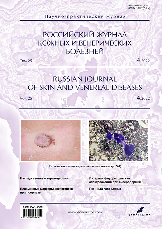
Amelanotic nodular malignant melanoma in a patient with a family history of skin cancers
摘要
Amelanotic skin melanoma is the amelanotic or hypomelanotic subtype of nodular melanoma. Amelanotic melanoma accounts for almost 60% of completely unrecognized melanoma. Amelanototic melanomas may not have the classic features of other melanomas and are usually red or skin-colored and more symmetrical, so superficially spreading melanomas are more easily recognized by most healthcare professionals and even patients.
Today, the path of melanoma development associated with genetic predisposition is being considered, in particular, with an unfavorable oncological family history, which is characterized by the development of melanoma in areas of the body periodically exposed to insolation, such as the trunk and limbs. The mechanism underlying amelanosis is not fully understood. As in pigment analogs, amelanotic melanoma cells retain their melanin-producing ability due to the expression of tyrosinase and microphthalmia-associated transcription factor.
We present our own clinical case of the development of a rare amelanocytic nodular melanoma on the leg in a patient with a family history of cancer. The complexity of the presented clinical case lies in the clinical picture atypical for nodular melanoma, since the formation was not pigmented, which did not cause concern for the patient and doctors, since the clinical picture was more consistent with a typical pyogenic granuloma. The patient underwent a wide excision of the formation. Further examination ruled out tumor metastases. The patient received a course of prophylactic therapy with recombinant interferon alfa. There was no recurrence of the disease during 3 years of follow-up. No metastasis has been observed so far.
 261-268


DERMATOLOGY
Efficacy of NB-UVB and azathioprine combination in the therapy of nonsegmental vitiligo
摘要
BACKGROUND: Vitiligo is a multifactorial acquired disease characterized by the appearance of depigmented, clearly delineated patches, on various areas on the skin. Narrowband Ultraviolet B Therapy is a first-line therapy for nonsegmental vitiligo. At the same time, it often takes at least 6 months to achieve optimal results, which is not always convenient for patients, brings additional financial costs of treatment. Therefore, in most cases to reduce the number of phototherapy sessions requires the use of additional drugs aimed at stopping the progression of the process and with a minimal spectrum of side effects.
AIM: to compare the effectiveness of azathioprine in combination with NB-UVB and NB-UVB monotherapy in progressive non-segmental vitiligo.
MATERIALS AND METHODS: The study included 60 patients with advanced non-segmental vitiligodivided into two groups of 30 people in each. Group A patients received therapy with azathioprine in combination with NB-UVB, and Group B patients ― NB-UVB monotherapy. VASI (Vitiligo Area Scoring Index) and DLQI (Dermatology Life Quality Index) were used for all patients to evaluate therapy efficacy.
RESULTS: All 60 patients diagnosed with nonsegmental progressive vitiligo completed the study and were included in the final analysis. Among patients who received the combined therapy protocol of azathioprine combined with narrowband phototherapy, a statistically more significant reduction in the severity and prevalence of the skin process was noted compared to the group of patients who received NB-UVB alone. More significant arresting of disease progression was noticed: in group A, among 30 patients, only 4 patients developed new lesions of vitiligo within 6 months of therapy, and in group B ― in 11 out of 30 patients. Group A patients showed a more significant reduction of VASI and DLQI compared to the control group.
CONCLUSION: Thus, the combination of NB-UVB and azathioprine in the treatment of non-segmental vitiligo showed great efficacy in arresting and reducing of the disease activity, prevalence and severity of vitiligo. Azathioprine is well tolerated by patients and has a low spectrum of side effects, which allows its successful use to stabilize vitiligo and stimulate repigmentation of foci.
269-278
Effect of phototherapy on psychoemotional state in psoriasis: assessment of depression, anxiety and quality of life
摘要
BACKGROUND: Psoriasis is a widespread chronic genetically determined inflammatory dermatosis that affects approximately 125 million people worldwide. In the vast majority of cases, the first rashes appear in people younger than 35 years old who are just starting to build a career and start a family. Localization of psoriatic rashes on visible areas of the skin and stigmatization in society, regular exacerbations and multiple courses of expensive and insufficiently effective treatment negatively affect the quality of life and emotional state of patients up to the development of depressive and anxiety disorders, and even suicide attempts. An imbalance of mental status leads to the progression of psoriasis, which further aggravates the course of the skin process. This necessitates a personalized approach to the choice of a therapy method, taking into account high efficiency and maximum comfort for patients, which will improve their psychoemotional state.
AIM: to study the effect of phototherapy (PUVA and UVB-311 nm) on the mental status of psoriasis patients
MATERIALS AND METHODS: A prospective study was conducted on the basis of FGKU “Central Polyclinic” and V.A. Rakhmanov’s clinic of skin and venereal diseases of Sechenov University with the participation of 228 patients with moderate-to-severe psoriasis who received UVB-311 nm (n=116) and PUVA therapy (n=112), including psoriasis vulgaris (n=193), guttate psoriasis (n=14), exudative psoriasis (n=21). Before and after treatment, patients were assessed for the severity of psoriasis (PASI), quality of life (DQLI), as well as the presence of depression and anxiety disorders (panic disorder, social phobia) using PHQ-9 and GAD-7 questionnaire tests. The initial radiation dose was 1 J/cm2 at PUVA and 0.1–0.2 J/cm2 at UVB-311 nm with a gradual increase in the dose every 1–2 sessions by 0.5 J/cm2 and 0.1 J/cm2, respectively. As photosensitizers ammifurin and oxoralen were used. The treatment was carried out according to the method of four-time irradiation per week using the UV-7001K cabin (Waldmann, Germany).
RESULTS: Among 228 patients there were 108 women and 120 men aged 43±8.2 years. The UVB-311 nm group included 102 patients with moderate psoriasis vulgaris and 14 – guttate psoriasis, the second group (PUVA) included patients with severe recalcitrant psoriasis vulgaris (n=91) and exudative psoriasis (n=21). In the first group, the course of therapy consisted of 28±2 procedures of UVB-311 nm. Before treatment, minimal depression was observed in 19 (16%) patients, mild depression – in 87 (75%), moderate depression – in 10 (9%). A high level of anxiety was observed in 94 (81%), the average level – in 22 (19%) patients. At the end of the course of UVB, PASI 90 was achieved in 94% (n=109) of patients, PASI 100 – in 88% (n=102); at the same time, on average, anxiety decreased by 80%, depressive symptoms decreased by 73%, quality of life increased by 92%. In the second group, the PUVA course consisted of 25±3 irradiation sessions. Before starting therapy, minimal depression was observed in 12 (11%) cases, mild depression – in 73 (65%), moderate depression – in 27 (24%). A high level of anxiety was characteristic of 100% of patients. As a result of therapy, PASI 90 was achieved in 95.5% (n=107) of cases, PASI 100 – in 90% (n=101). The average severity of anxiety, depression, and quality of life improved by 88%, 83%, and 96% respectively.
CONCLUSION: PUVA and UVB-311 nm have demonstrated high efficacy and safety in moderate-to -severe psoriasis, contributing to the rapid cleansing of the skin from rashes and improving the psycho-emotional state, which not only significantly improves the quality, but also increases the life expectancy of patients.
.
279-287
The place of laser fluorescence spectroscopy, doppler flowmetry and ultrasound in the diagnosis and assessment of treatment efficacy for plaque scleroderma
摘要
According to modern ideas, a reasonable choice of an effective method of treating plaque scleroderma is based on the diagnosis of the pathological process prevailing in the tissues (inflammation-sclerosis). Therefore, an urgent problem of a personalized approach to dermatosis therapy is the possibility of an objective assessment of the prevailing process using non-invasive diagnostic methods. The article presents a clinical case of widespread plaque scleroderma in a 66-year-old patient, demonstrating the possibility of using laser fluorescence spectroscopy and laser Doppler flowmetry to determine the degree of activity of the focus and determine the leading pathological process.
We selected three pathological skin foci localized in the abdomen and characterizing three clinical stages of the disease (inflammation, induration, sclerosis). The analysis of fluorescence and laser Doppler flowmetry data showed that in areas clinically defined as inflammation, there is an increase in the average values of the indices of tissue content of porphyrins, lipofuscin and microcirculation index compared with intact skin, while the intensity of collagen fluorescence does not differ significantly. In the induration zone, along with an increase in the fluorescence indices of lipofuscin and porphyrins, there is an increase in the average values of collagen fluorescence indices at effective registration waves. The data obtained by us indicate an active inflammatory process in these foci and the process of fibrosis in the induration zone. In the sclerosis zone, there is an increase in the average values of collagen fluorescence indices compared with intact skin, and the fluorescence of optical markers of inflammation (lipofuscin and porphyrins) do not differ significantly in comparison with the control intact skin. When analyzing the fluorescence spectra and laser Doppler flowmetry data after treatment, we found that in the zones of induration and inflammation, the average values of the fluorescence indices of porphyrins, lipofuscin, collagen and microcirculation index are reduced relative to the initial values (before treatment), but remain higher in comparison with intact skin. The data obtained may indicate that active inflammation in these foci persists at the time of the study. In the study of the focus of sclerosis, the obtained autofluorescence and microcirculation data in dynamics do not differ significantly from the initial values. The data for laser Doppler flowmetry and laser fluorescence spectroscopy are consistent with ultrasound examination of the skin.
In our study, the potential possibility of using laser fluorescence spectroscopy and laser Doppler flowmetry methods to establish the degree of activity of the focus, determine the leading pathological process, as well as to evaluate the effectiveness of therapy was demonstrated for the first time.
289-302
Hereditary keratoderma. Clinical case: Unna–Toast keratoderma and Buschke–Fischer–Brauer keratoderma
摘要
Palmoplantar keratoderma is a heterogeneous group of diseases, both hereditary and acquired, affecting, as a rule, the skin of the palms and soles in the form of focal or diffuse hyperkeratotic layers. Hereditary forms are characterized by a defect in the genes encoding certain structural components of keratinocytes, which leads to the corresponding clinical manifestations. The actual method of genomic sequencing has made it possible to identify the localization of defects in certain genes for specific types of hereditary palmoplantar keratoderma, however, despite the big step in diagnosis, therapy still remains complex and can only reduce the manifestations of the disease and improve the quality of life, and not cure it completely.
The authors demonstrate a clinical case of Unna–Toast keratoderma in a young patient with a long history of treatment of the disease, who showed a pronounced positive dynamics from the skin process against the background of the use of local agents, as well as the use of such a physiotherapeutic method as local PUVA therapy. In addition, the article provides a clinical case of hereditary keratoderma Buschke–Fischer–Brauer as an example of an accidentally diagnosed disease at a dermatologist’s appointment.
303-312
Widespread dermatophytosis after combined immunosuppressive therapy in a patient with COVID-19
摘要
Mycotic diseases of human skin are frequently diagnosed reaching 20–25% of the population prevalence. The leading factor in the mycotic prevalence is the high survival rate of fungi in various environmental conditions. Up to 30 genera of fungi constantly live on human skin and mucous membranes. This constant balance of their presence on the body and interaction with it is called a mycobiome. If the balance between it and the macroorganism is disturbed, the reproduction of fungi begins and mycotic skin lesions develop.
The study included the patient V. 75 years old, who came in with complaints of widespread rashes on the skin of the body and limbs accompanied by peeling after a COVID-19 infection. Microscopy revealed mycelium of pathogenic fungi. The patient received systemic and topical antifungal therapy until the end of hospitalization. He continued to receive terbinafine on an outpatient basis in combination with a topical antimycotic after discharge. Short review on baricitinib and levilimab is presented. According to the results of the study, a 60–70% regression of the skin process was achieved after 2 weeks at discharge. One month later a complete regression of the skin process was achieved due to the therapy.
The use of the combination of janus-kinase inhibitors with biological drugs and systemic glucocorticosteroids in COVID-19 patients has a powerful immunosuppressive effect on the body. It leads to the development of a common fungal skin lesion, which was observed in our patient.
313-321
The role of Vitamin D in the pathogenesis of some immune-mediated dermatoses
摘要
In recent years, the sunlight vitamin has become extremely popular and almost mandatory to use, especially because of its pleiotropic effects, although until recently its use was limited to the prevention of the development of pathology of the bone system, in particular rickets in children. Being actually a fat-soluble prohormone of a steroid nature, Vitamin D participates in the endocrine, paracrine and autocrine regulation of the body. The pharmacotherapeutic renaissance of calciferol is associated with the discovery of Vitamin D receptors in most cells of the body, and the presence of enzymes synthesizing the active form of Vitamin D extrarenally, in particular, in the skin, has led to renewed interest and broad discussion in the dermatological community. Is the role of non-bone effects of calciferol, mainly its role in the pathogenesis of autoimmune skin diseases, really justified from the point of view of evidence-based medicine, and is the tendency to consume Vitamin D safe?
This article presents the most up-to-date information about the role of Vitamin D deficiency in the mechanisms of immune response development, in some dermatoses. In addition to generalizing the bone and extra-bone functions of Vitamin D to the macroorganism, the mechanisms of formation of some of the most common dermatoses, such as psoriasis, atopic dermatitis and vitiligo are discussed in detail. The review details the biological effects of Vitamin D in the skin. The article analyzes the legality of the use of Vitamin D-based drugs and their effectiveness in dermatological practice.
323-332
CHRONICLES
Chronicles of A.I. Pospelov Moscow Dermatovenerology and Cosmetology Society (MDCS was founded on October 4, 1891) Bulletin of the MSDC № 1140
摘要
On November 17, 2020, the 1140th meeting was held in on-line format.
In total there were 92 participants. Five people were accepted as members of the MDCS.
The clinical agenda of the meeting considered cases of subacute (disseminated) cutaneous lupus erythematosus and reactions to tattooing in the form of pseudolymphoma of the skin. The interest of observing disseminated lupus erythematosus was in the difficulties of diagnosis due to its combination with Sjogren’s syndrome, a long history of the disease, the severity of the skin process and the peculiarities of the selection of adequate drug therapy. Analysis of the case of pseudolymphoma development as a reaction to a tattoo in a patient with atopic dermatitis helped to approach a more accurate understanding of the clinical and pathogenetic aspects of this process: probably, the pathophysiological reaction is based on a reaction to a foreign body, which makes this disease related to sarcoidosis.
The scientific agenda of the meeting reported on the advantages of using photodynamic therapy for skin T-cell lymphomas, such as good cosmetic results; non-invasive nature of the procedure; selectivity; low risk of toxicity; low photosensitivity; low carcinogenic potential. In another report ― on the use of signaling pathway blockers in the treatment of psoriasis ― the results of an open uncontrolled prospective clinical study of the effectiveness of apremilast in the treatment of moderate and severe psoriasis were presented.
333-336
PHOTO GALLERY
Photogallery. Hidradenitis suppurativa (acne inversa)
摘要
Hidradenitis suppurativa (acne inversa) is a chronic, inflammatory, primary follicular disease triggered by follicular occlusion with subsequent inflammation and destruction of the skin appendage, affecting hair follicles located in apocrine gland-bearing body areas. Clinical presentation of hidradenitis suppurativa is extremely variable showing a wide spectrum of cutaneous lesions in different stages of evolution, different pattern of distribution and grades of severity. Traditionally for severity staging the Hurley clinical grading system has been used where stage I stands for mild disease while stage II and III for moderate and severe stages respectively. 2015 classification of Van Der Zee and Jemec proposed 6 phenotypes of hidradenitis suppurativa: regular type, frictional furuncle type, scarring folliculitis type, conglobata type, syndromic type, ectopic type.
We present a photogallery on this problem.
337-340
Регистрационный номер и дата принятия решения о регистрации СМИ: серия ПИ № ФС 77 - 86501 от 11.12.2023 г.
СМИ зарегистрировано Федеральной службой по надзору в сфере связи, информационных технологий и массовых коммуникаций (Роскомнадзор).
Регистрационный номер и дата принятия решения о регистрации СМИ: серия ЭЛ № ФС 77 - 80653 от 15.03.2021 г.