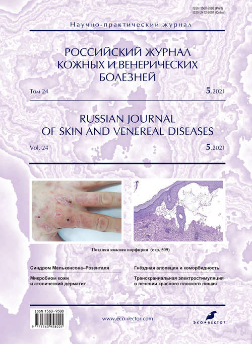
Clinical case of porphyria cutanea tarda
- 作者: Motorina A.V.1, Palkina N.V.1, Khorzhevsky V.A.1, Fefelova Y.A.1
-
隶属关系:
- Professor V.F. Voino-Yasenetsky Krasnoyarsk State Medical University
- 期: 卷 24, 编号 5 (2021)
- 页面: 503-508
- 栏目: CLINICAL PICTURE, DIAGNOSIS, AND THERAPY OF DERMATOSES
- ##submission.dateSubmitted##: 16.01.2022
- ##submission.dateAccepted##: 14.02.2022
- ##submission.datePublished##: 12.09.2021
- URL: https://rjsvd.com/1560-9588/article/view/96529
- DOI: https://doi.org/10.17816/dv96529
- ID: 96529
如何引用文章
全文:
详细
Cutaneous porphyria tarda is a rare skin disease characterized by a chronic course and the appearance of painful vesicular elements on the skin, localized mainly in open areas of the body under the influence of ultraviolet radiation. It is known that the development of porphyria in about a third of cases is familial and can also be associated with alcohol abuse, exposure to gasoline and other toxic substances, smoking, the presence of HIV infection, high iron levels in the body and liver damage with viral hepatitis C. A key link in the pathogenesis late cutaneous porphyria is considered a violation of the function of one of the enzymes in the liver cells involved in the synthesis of heme – uroporphyrinogen decarboxylase, which causes a violation of pigment metabolism with increased accumulation of porphyrins in the body, which are deposited in the skin and act as endogenous photosensitizers.
As an auxiliary tool for differential diagnosis in case of suspicion of late cutaneous porphyria, in addition to collecting a detailed anamnesis, it is necessary to clarify the presence of a pathognomonic symptom ― a change in the color of urine, since the accumulation of porphyrin in the body in some cases can change the color of urine to reddish-brown. Another tool is a skin biopsy, which usually helps with differential diagnosis.
In the treatment of late cutaneous porphyria, in addition to the treatment of viral hepatitis C, low doses of hydroxychloroquine are used. Although the disease is not completely curable, its course can be successfully controlled. In this regard, timely diagnosis of tardive cutaneous porphyria allows for control of the disease. The elimination and treatment of skin manifestations consists in the primary elimination of those factors that provoked the exacerbation of the disease. Such patients need to wear protective clothing, limit exposure to sunlight as much as possible, since to date there are no means that significantly reduce the level of porphyrin and restore the enzymatic insufficiency of uroporphyrinohendecarboxylase.
Molecular genetic testing is relevant in order to further reduce the risk of developing the disease in offspring, which requires limiting factors that reduce the levels and activity of uroporphyrinohendecarboxylase.
The search and development of effective drugs for the treatment of late cutaneous porphyria continues all over the world.
In the present paper we present a clinical case of cutaneous porphyria tarda associated with hepatitis C virus infection.
全文:
ВВЕДЕНИЕ
Современные исследования показывают, что развитие заболевания поздней кожной порфирии зачастую связано с печёночной недостаточностью, а именно с нарушением активности печёночного фермента уропорфириногендекарбоксилазы.
Начало заболевания регистрируется в довольно зрелом возрасте. Мужской пол, наличие вирусного гепатита C, ВИЧ-инфекции, злоупотребление алкоголем, избыток железа в организме повышают риск развития заболевания, так как указанные факторы напрямую снижают уровень уропорфириногендекарбоксилазы [1]. Данный фермент превращает уропорфириноген III в копропорфириноген III, что является одним из этапов синтеза гема [2]. Недостаточность фермента приводит к накоплению в коже производных порфирина, которые связываются с белком TSPO, опосредующим антиоксидантные процессы [3].
При окислении производных порфирина под воздействием ультрафиолетового излучения вызывается активация мастоцитов и их дегрануляция с формированием специфичных для этого заболевания пузырей на открытых участках кожи [4]. Другим специфичным изменением при поздней кожной порфирии является развитие гипертрихоза [5]. Сравнительно необычное осложнение заболевания ― развитие псевдосклеродермии с характерными изменениями компонентов внеклеточного матрикса, в частности увеличением содержания коллагена в коже престернальной области [6, 7].
Терапия данного заболевания заключается в назначении низких доз гидроксихлорохина, применении солнцезащитных средств, терапии вирусного гепатита С.
ОПИСАНИЕ КЛИНИЧЕСКОГО СЛУЧАЯ
О пациенте
Пациент Ч., 1980 года рождения, в июне 2021 года обратился к дерматологу с жалобами на пузыри на кожных покровах, долго заживающих на открытых участках тела.
При осмотре: на открытых участках тела, а именно кистях рук, лице, шее имеются множественные везикулы и буллы с признаками атрофии, гипер- и гипопигментации (рис. 1). Со слов больного, с 2011 года страдает вирусным гепатитом C.
Рис. 1. Поражение кожи кистей рук. / Fig. 1. Skin lesions of the hands.
На основании анамнеза, клинических проявлений и лабораторных исследований, а также результатов патоморфологического исследования пациенту выставлен диагноз: «Поздняя кожная порфирия». Рекомендовано наблюдение гепатолога и терапевта.
Результаты физикального, лабораторного и инструментального исследования. При постановке реакции иммунофлюоресценции определяется линейное отложение IgG, IgM вдоль базальной мембраны. В реакциях с антителами IgA свечения нет (рис. 2). При гистохимическом исследовании обнаружены субэпидермальные пузыри, состоящие из малодифференцированных фибробластов; в верхней части дермы и утолщённых стенках капилляров имеются отложения гиалина (рис. 3). В стенках сосудов выявлены ШИК*-положительные диастазорезистентные вещества (рис. 4).
Рис. 2. Линейное отложение IgG и IgM вдоль базальной мембраны; IgA ― свечения нет. ×50. / Fig. 2. Linear deposition of IgG, IgM along the basement membrane; IgA ― no luminescence. ×50.
Рис. 3. Результаты гистохимического исследования кожи: отложения гиалина в верхней части дермы и утолщённых стенках капилляров. ×50. / Fig. 3. Results of histochemical assay of the skin: hyaline deposits in the upper part of the dermis and thickened capillary walls. ×50.
Рис. 4. ШИК-реакция с диастазой: в стенках сосудов выявлены ШИК-положительные диастазорезистентные вещества. ×100. / Fig. 4. Periodic acid-Schiff reaction with diastasе: PAS-positive diastase-resistant substances were detected in the vessel walls. ×100.
Дифференциальный диагноз. Данная патология требует дифференциальной диагностики с буллёзным эпидермолизом, экземой, буллёзной формой многоформной экссудативной эритемы, системной склеродермией. В данном случае решающим является результат исследования на порфирины.
Анализ мочи на общие порфирины положительный.
Исход и результаты последующего наблюдения. О дальнейшем состоянии пациента информации нет.
ОБСУЖДЕНИЕ
В качестве вспомогательного инструмента дифференциальной диагностики при подозрении на позднюю кожную порфирию у пациента, кроме сбора подробного анамнеза с тщательным расспросом о любых факторах восприимчивости, обсуждаемых выше, таких как алкоголь, курение, воздействие химических веществ, следует уточнить наличие патогномоничного симптома, а именно изменение окраски мочи, поскольку накопление порфирина в организме в некоторых случаях может изменить цвет мочи на красновато-коричневый. Кроме того, мочу, кровь и стул пациента можно дополнительно исследовать на предмет выявления повышенного уровня порфирина [8].
Другим инструментом служит биопсия кожи, которая обычно не требуется для диагностики данного заболевания, но помогает при дифференциальной диагностике. При этом в литературных источниках сообщается, что результатом данного исследования при кожных порфириях, как правило, является гистологическая картина субэпидермальных пузырей и отложений иммунных комплексов [9], что соответствует гистологической картине результатов биопсии пациента в представленном клиническом случае.
Кроме общих рекомендаций, которыми являются избегание воздействия ультрафиолетового излучения (защитные кремы; ношение одежды, защищающей от воздействия ультрафиолетового излучения), ограничение попадания в организм токсических веществ, ограничение употребления алкоголя, данному больному показано назначение гидроксихлорохина [1]. Рекомендована консультация врача-инфекциониста для решения вопроса о терапии вирусного гепатита, так как прогноз заболевания поздней кожной порфирии осложняется наличием данного инфекционного заболевания. Важно отметить, что патогенез ухудшения течения поздней порфирии с сопутствующим гепатитом С связан не со снижением активности ферментов, а с прямым повреждением клеток печени [10].
Несмотря на то, что порфирии ― редкая группа метаболических нарушений, которые могут быть как унаследованными, так и приобретёнными, данное заболевание в настоящее время приобретает большое значение, так как охватывает многие социально-гигиенические вопросы: влияние на организм человека профессиональных вредностей, бытовых факторов, вредных привычек и т.д.
В связи с этим необходимо обсудить провоцирующее действие алкоголя на нарушенный порфириновый обмен, что даёт возможность правильно оценить роль этого вещества в патогенезе поздней кожной порфирии. Несмотря на то, что этиловый спирт не является истинным, а только лишь провоцирующим печёночным порфириногеном, заболевание порфирией у лиц, употребляющих алкоголь, протекает более тяжело. Это обусловлено также тем, что при употреблении алкогольных напитков происходит снижение уровня гепсидина и увеличивается всасывание железа [11].
ЗАКЛЮЧЕНИЕ
Устранение и лечение кожных проявлений заключается в первостепенном устранении тех факторов, которые спровоцировали обострение заболевания. Таким пациентам требуется ношение защитной одежды, ограничение воздействия солнечного света по мере возможности, так как на сегодняшний день средств, существенно снижающих уровень порфирина и восстанавливающих ферментативную недостаточность уропорфириногендекарбоксилазы, не существует.
Актуально молекулярно-генетическое тестирование с целью последующего снижения риска развития заболевания у потомства, для чего требуется ограничение факторов, снижающих уровни и активность уропорфириногендекарбоксилазы.
В отношении лечения поиск и разработка эффективных препаратов продолжается во всём мире.
ДОПОЛНИТЕЛЬНО
Источник финансирования. Авторы заявляют об отсутствии внешнего финансирования при подготовке статьи.
Конфликт интересов. Авторы декларируют отсутствие явных и потенциальных конфликтов интересов, связанных с публикацией настоящей статьи.
Вклад авторов. А.В. Моторина ― анализ данных и интерпретация результата, написание статьи; Н.В. Палкина, В.А. Хоржевский ― получение, анализ данных и интерпретация результата, внесение в рукопись важной правки с целью повышения научной ценности статьи; Ю.А. Фефелова ― интерпретация данных, внесение в рукопись важной правки. Все авторы подтверждают соответствие своего авторства международным критериям ICMJE (разработка концепции, подготовка работы, одобрение финальной версии перед публикацией).
Согласие пациента. Пациент добровольно подписал информированное согласие на публикацию персональной медицинской информации в обезличенной форме в журнале «Российский журнал кожных и венерических болезней».
ADDITIONAL INFORMATION
Funding source. This work was not supported by any external sources of funding.
Competing interests. The authors declare that they have no competing interests.
Author contribution. A.V. Motorina ― data analysis and interpretation of the result, writing an article; N.V. Palkina, V.A. Khorzhevsky ― obtaining, analyzing data and interpreting the result, making an important edit to the manuscript in order to increase the scientific value of the article; Yu.A. Fefelova ― a significant contribution to the interpretation of the data, making an important revision to the manuscript. The authors made a substantial contribution to the conception of the work, acquisition, analysis of literature, drafting and revising the work, final approval of the version to be published and agree to be accountable for all aspects of the work.
Patients permission. The patient voluntarily signed an informed consent to the publication of personal medical information in depersonalized form in the journal “Russian journal of skin and venereal diseases”.
* Реакция Шифф-йодной кислотой ― кожная проба, предложенная австрийским педиатром B. Schick (1877–1967), применяемая для оценки выраженности местной реакции.
作者简介
Anna Motorina
Professor V.F. Voino-Yasenetsky Krasnoyarsk State Medical University
Email: kozlovaa.v@mail.ru
ORCID iD: 0000-0003-2211-1708
SPIN 代码: 4368-3288
MD, Cand. Sci. (Med.)
俄罗斯联邦, 1, Partizanа Zheleznyakа str., Krasnoyarsk, 660022Nadezhda Palkina
Professor V.F. Voino-Yasenetsky Krasnoyarsk State Medical University
编辑信件的主要联系方式.
Email: MosmanNV@yandex.ru
ORCID iD: 0000-0002-6801-3452
SPIN 代码: 7534-4443
Scopus 作者 ID: 56126629300
MD, Cand. Sci. (Med.)
俄罗斯联邦, 1, Partizanа Zheleznyakа str., Krasnoyarsk, 660022Vladimir Khorzhevsky
Professor V.F. Voino-Yasenetsky Krasnoyarsk State Medical University
Email: vladpatholog@yandex.ru
ORCID iD: 0000-0002-9196-7246
MD, Cand. Sci. (Med.)
俄罗斯联邦, 1, Partizanа Zheleznyakа str., Krasnoyarsk, 660022Yulia Fefelova
Professor V.F. Voino-Yasenetsky Krasnoyarsk State Medical University
Email: fefelovaja@mail.ru
ORCID iD: 0000-0001-5434-7155
SPIN 代码: 9210-6780
Dr. Sci. (Biol.), Associate Professor
俄罗斯联邦, 1, Partizanа Zheleznyakа str., Krasnoyarsk, 660022参考
- Singal AK. Porphyria cutanea tarda: Recent update. Mol Gen Metab. 2019;128(3):271–281. doi: 10.1016/j.ymgme.2019.01.004
- Christiansen AL, Aagaard L, Krag A, et al. Cutaneous porphyrias: causes, symptoms, treatments and the danish incidence 1989–2013. Acta Derm Venereol. 2016;96(7):868–872. doi: 10.2340/00015555-2444
- Ruksha T, Aksenenko M, Papadopoulos V. Role of Translocator protein in melanoma growth and progression. Arch Derm Res. 2012;304(10):839–845. doi: 10.1007/s00403-012-1294-5.
- Lim HW. Mechanisms of phototoxicity in porphyria cutanea tarda and erythropoietic protoporphyria. Immunol Ser. 1989;46(7):671–685.
- Frank J, Poblete-Gutiérrez P. Porphyria cutanea tarda--when skin meets liver. Best Pract Res Clin Gastroenterol. 2010;24(5):735–745. doi: 10.1016/j.bpg.2010.07.002
- Ruksha TG, Aksenenko MB, Klimina GM, Novikova LV. Extracellular matrix of the skin: role in the development of dermatological diseases. Vestnik Dermatologii i Venerologii. 2013;89(6):32–39. (In Russ).
- Wallaeys E, Thierling U, Lang E, et al. Porphyria cutanea tarda with sclerodermatous changes and hemochromatosis. Hautarzt. 2014;65(4):272–274. doi: 10.1007/s00105-014-2783-6
- Stölzel U, Doss M, Schuppan D. Clinical guide and update on porphyrias. Gastroenterology. 2019;157(2):365–381.e4. doi: 10.1053/j.gastro.2019.04.050
- Maynard B, Peters MS. Histologic and immunofluorescence study of cutaneous porphyrias. J Cut Pathol. 1992;19(1):40–47. doi: 10.1111/j.1600-0560.1992.tb01557.x
- Moran MJ, Fontanellas A, Brudieux E, et al. Hepatic uroporphyrinogen decarboxylase activity in porphyria cutanea tarda patients: the influence of virus C infection. Hepatology (Baltimore, Md.). 1998;27(2):584–589. doi: 10.1002/hep.510270237
- Doss MO, Kühnel A, Gross U. Alcohol and porphyrin metabolism. Alcohol and Alcoholism (Oxford, Oxfordshire). 2000;35(2):109–125. doi: 10.1093/alcalc/35.2.109
补充文件


