Features of topical therapy of skin and hair mycosis caused by Microsporum genus fungi: Results of clinical observations
- 作者: Yakovlev A.B.1
-
隶属关系:
- Central State Medical Academy of Department of Presidential Affairs
- 期: 卷 25, 编号 5 (2022)
- 页面: 381-388
- 栏目: DERMATOLOGY
- ##submission.dateSubmitted##: 19.10.2022
- ##submission.dateAccepted##: 09.11.2022
- ##submission.datePublished##: 11.12.2022
- URL: https://rjsvd.com/1560-9588/article/view/111992
- DOI: https://doi.org/10.17816/dv111992
- ID: 111992

如何引用文章

详细
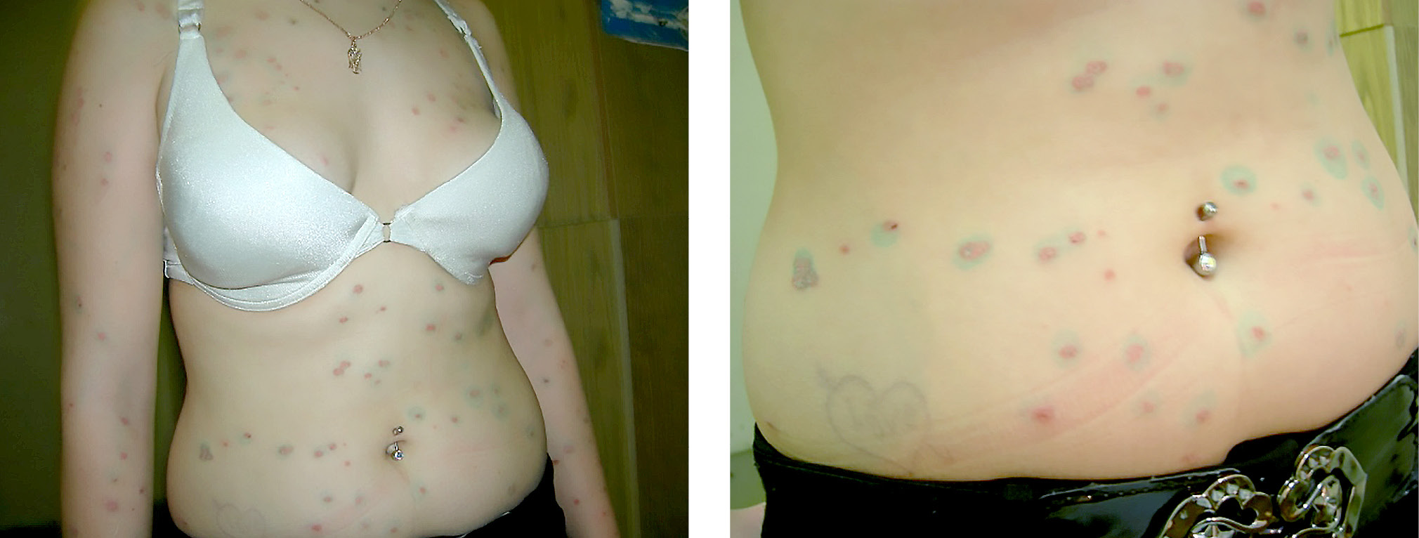
The article summarizes the long-term experience of observing the dynamics of pathological process of tinea corporis caused by Microsporum spp. in children and adults treated at the initial stage with combined topical steroids and without them. The duration of treatment increases by 1.5–2 times when combined topical steroids are used, while with other mycoses caused by fungi of the genera Trichophyton, Epidermophyton, this does not occur. This can be explained, apparently, by the peculiarities of sporulation of fungi of the genus Microsporum spp.: most dermatomycetes at the time of penetration into the epidermis and dermis rely on the germination of mycelium, and only in fungi of the genus Microsporum spp. at the time of invasion, sporulation activity does not decrease. As a result, the fungus is able to simultaneously infect the epidermis and hair.
Most modern therapeutic programs for the external treatment of microsporia include iodine preparation and an antimycotic in the form of a cream or ointment. These drugs do not suppress the macrophage reaction in the lesion, and with the onset of treatment, sporulation decreases sharply. When using a topical combination drug containing an antimycotic and combined topical steroids, the fungus continues to actively sporulate, as a result of which the process in the epidermis stops, and the spores that have penetrated into the hair remain viable.
全文:
作者简介
Alexey Yakovlev
Central State Medical Academy of Department of Presidential Affairs
编辑信件的主要联系方式.
Email: ale64080530@yandex.ru
ORCID iD: 0000-0001-7073-9511
SPIN 代码: 6404-7701
MD, Cand. Sci. (Med.), Associate Professor
俄罗斯联邦, 19 Marshala Timoshenko str., Moscow, 121359参考
- Sergeev AY, Sergeev YV. Fungal infections. A guide for doctors. 2nd ed. Moscow: BINOM; 2008. 480 p. (In Russ).
- Federal clinical guidelines. Dermatovenereology. 2015. Skin diseases. Sexually transmitted infections. 5th ed., revised and updated. Moscow: Business Express; 2016. 768 p. (In Russ).
- Garibova LV, Lekomtseva SN. Fundamentals of mycology. Morphology and systematics of fungi and mushroom-like organisms. Textbook. Moscow: Association of scientific publications of the CMC; 2005. 220 p. (In Russ).
- Rodionov AN, Zaslavsky DV, Sydikov AA. Dermatology. Illustrated guide to clinical diagnostics. Ed. by A.N. Rodionov. Moscow: Border; 2018. 944 p. (In Russ).
- Calander S, Saunte DM, Polesie S. Tinea capitis caused by microsporum audouinii: lessons from a Swedish community outbreak. Acta Dermatol Venereol. 2021;101(9):adv00551. doi: 10.2340/00015555-3909
- Serebryakova IS, Kornisheva VG, Ravodin RA, et al. Skin mycosis caused by Nannizzia incurvata (formerly Microsporum incurvatum): Description of a rare clinical special case. Problems Med Mycology. 2019;21(1):16−20. (In Russ).
- Sergeev AY, Burova SA, Kasikhina EI. Dermatomycoses in the era of pandemic. Immunopathol, Allergol, Infectol. 2021;(1)79−96. (In Russ). doi: 10.14427/jipai.2021.1.79
- Sybren de Hoog G, Dukik K, Monod M, et al. Toward a novel multilocus phylogenetic taxonomy for dermatophytes. Mycopathologia. 2017;182(1-2):5–31. doi: 10.1007/s11046-016-0073-9
- Klimko NN. Mycoses: diagnosis and treatment. Guide for doctors. Moscow: Premier MT; 2007. 336 p. (In Russ).
- Yakovlev AB. Microsporia. Trichophytosis. Favus. A textbook for doctors. 2nd ed., revised. Moscow: Novik; 2014. 140 p. (In Russ).
- Lavrushko SI, Stepanenko VI. Modern diagnosis and complex treatment of microsporia in athletes. East Eur Sci J. 2021;2(8):9−15. (In Russ). doi: 10.31618/ESSA.2782-1994.2021.2.72.112
- Kassem R, Shemesh Y, Nitzan O, et al. Tinea capitis in an immigrant pediatric community: A clinical signs-based treatment approach. BMC Pediatrics. 2021;21(1):363. doi: 10.1186/s12887-021-02813-x
- Tikhonovskaya IV, Adaskevich VP, Shafranskaya TV. Microsporia in children: Clinic, diagnosis and treatment. Recipe. 2006;(3):72−74. (In Russ).
- Dermatovenerology. National leadership. Short Edition. Ed. by Y.S. Butov, Y.K. Skripkin, O.L. Ivanov. Moscow: GEOTAR-Media; 2017. 896 p. (In Russ).
- Medvedeva TV, Chilina GA. The case of isolation of a rare causative agent of microsporia. Adv Med Mycology. 2015;(14):34. (In Russ).
- Kulaga VV, Romanenko IM, Safonov SL, Kulaga SM. Fungal diseases and their complications. Guide for doctors. Moscow: Medical Information Agency; 2010. 688 p. (In Russ).
- John AM, Schwartz RA, Janniger CK. The kerion: an angry tinea capitis. Int J Dermatol. 2018;57(1):3–9. doi: 10.1111/ijd.13423
- Abidova ZM, Rakhimov IR, Karabaeva IT. Characteristics of cytokine disorders in patients with microsporia. Adv Med Mycology. 2018;(18):285–286. (In Russ).
- Antonova SB, Ufimtseva MA, Bochkarev YM. Atypical micro-sporia: A “transformed” variant. A case from practice. Contemporary Problems Sci Education. 2015;(5):306–314. (In Russ).
- Tarasenko GN, Tarasenko YG. Smooth skin mycoses: approaches to diagnosis and therapy. Hospital Medicine: Sci Practice. 2020;(3):32–36. (In Russ).
- Le TK, Cohen BA. Tinea capitis: advances and a needed paradigm shift. Curr Opinion Pediatr. 2021;33(4):387–391. doi: 10.1097/MOP.0000000000001034
- Lavrushko SI. Complex treatment of microsporia of the scalp in children. Ukr J Dermatol Venereol Cosmetol. 2019;(1):65–72. (In Russ).
补充文件