


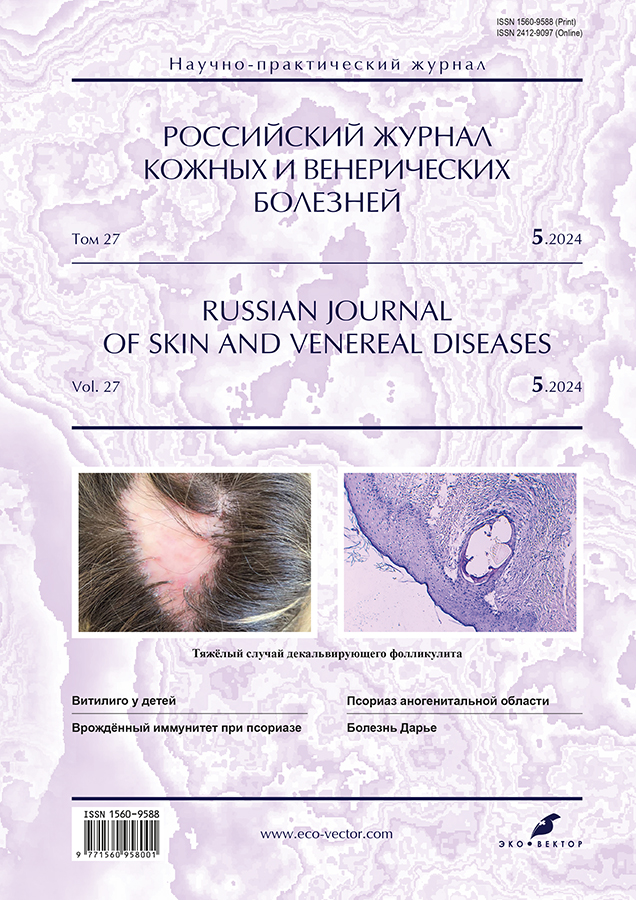
A case of severe folliculitis decalvans
- Authors: Teplyuk N.P.1, Pinegin V.B.1, Varshavsky V.A.1, Brezhneva A.A.1
-
Affiliations:
- I.M. Sechenov First Moscow State Medical University (Sechenov University)
- Issue: Vol 27, No 5 (2024)
- Pages: 526-537
- Section: DERMATOLOGY
- Submitted: 16.07.2024
- Accepted: 15.09.2024
- Published: 01.12.2024
- URL: https://rjsvd.com/1560-9588/article/view/634377
- DOI: https://doi.org/10.17816/dv634377
- ID: 634377

Cite item

Abstract
Folliculitis decalvans is a difficult to treat rare neutrophilic dermatosis that affects the scalp and leads to permanent scarring. The pathogenesis is currently not well understood; the influence of Staphylococcus aureus antigens, a disorder of the skin microbiome, a defect in cell-mediated immunity, and an imbalance of pro-inflammatory cytokines are assumed. This disease is often associated with a marked decrease in quality of life, because in addition to significant external manifestations, the patient may experience pain, itching and burning in the areas of the rash. The main problem is that there is currently no effective therapy for this disease. Treatment options include systemic antibiotics, topical and intralesional corticosteroids, dapsone, isotretinoin, biologics, and photodynamic therapy. Administration of radiation therapy, adipose tissue transplantation, use of excimer laser, and intravenous injections of human immunoglobulin have also been reported.
Decalving folliculitis is a disease that requires the earliest possible diagnosis, as its outcome is persistent scarring atrophy of the hair follicle, marked discomfort of the patient, constant sensation of soreness and burning. It is necessary to use the most effective therapy, change the treatment strategy if the disease continues to progress.
We present a brief overview of the main methods of therapy and a clinical case of a long course of severe folliculitis decalvans, refractory to the main methods of treatment.
Full Text
Background
Folliculitis decalvans (FD) is a rare primary neutrophilic dermatosis that causes permanent scarring and complete destruction of hair follicles [1]. The condition was first described in 1888 by the French dermatologist Charles-Eugène Quinquaud [2]. FD accounts for 10.5% of cases of cicatricial alopecia and 2.8% of all cases of alopecia [3, 4]. The parietal scalp is the primary localization of lesions in 61.3% of cases [5]. However, some clinical reports describe lesions on limbs and trunk in addition to scalp involvement [6]. The literature shows that FD predominantly affects young and middle-aged individuals, with a slight male predominance [7]. Early onset before the age of 25 years is associated with more severe disease progression [1].
The pathogenesis remains poorly understood. Proposed mechanisms include hereditary immune dysregulation and staphylococcal superantigens [8]. Some evidence suggests that FD may be caused by disruption of the epidermal barrier, allowing subepidermal invasion by opportunistic microorganisms or transient flora [9]. Although Staphylococcus aureus is commonly found in lesions, its exact pathogenic role remains controversial. Scalp colonization by S. aureus, combined with hypersensitivity and cell-mediated immunity defects, is thought to cause severe inflammatory responses that destroy follicles [10]. Trüeb et al. [11] identified pathogenic bacterial biofilms in the infra infundibular (deeper) portions of human scalp follicles. Microbial antigens and/or superantigens can activate T lymphocytes and induce the release of various proinflammatory (e.g., interferon gamma, IFN-γ) and profibrotic (e.g., interleukin 4, IL-4) factors [12]. Inflammasomes and the IL-1β signaling pathway were shown to influence the innate immune response. A Th17 immune response is thought to be involved, associated with local expression of proinflammatory mediators such as tumor necrosis factor alpha (TNF-α) [13].
The diagnosis of FD is based on clinical manifestations, trichoscopy, and histology [5].
Clinical manifestations include perifollicular erythema, inflammation, scalp atrophy and scarring, crusting, scaling, pustules, papules, and typical follicular tufts consisting of 5–20 hairs [14–16]. Pustules are considered a marker of the severity and activity of FD [17]. In addition to visible external manifestations, patients often have trichodynia, pruritus, and burning sensation in the affected area [7]. Based on the size of the scar, FD is classified as stage I (<2 cm), stage II (2–5 cm), stage III (>5 cm) [18].
Early histopathological features include loss of sebaceous glands, intrafollicular acanthosis, and fibrosis involving the follicular infundibulum. Active lesions have a dense perifollicular neutrophilic infiltrate that transforms into a lymphoplasmacytic infiltrate located near the dermal part of the hair follicle. Other features include multiple hairs emerging from the same follicula (polytrichia), fused infundibua with severe inflammation in the upper portion of the follicle, follicular hyperkeratosis, hyperplasia of the interfollicular epidermis, atrophy of the follicular epithelium, plasma cells in the infiltrate, follicular microcysts, and atrophy of the sebaceous glands [13, 19, 20].
Trichoscopic features of FD progression include perifollicular erythema, milky red and ivory areas with loss of follicular orifices, follicular pustules, perifollicular hemorrhages, yellowish tubular scales, yellow crusts, and hair tufts. Well-defined, thin, branching vessels are considered a sign of improvement. The severity of FD depends on the extent of an alopecia scar [18, 21].
A folliculitis decalvans and lichen planopilaris phenotypic spectrum (FDLPPPS) combining clinical, trichoscopic and histopathological features of both diseases has been described [22, 23].
FD is one of the most difficult to treat scalp diseases, which has a significant impact on the patient’s life [13, 14, 24]. There is currently no effective treatment. Treatment failure often leads to further disease progression, increased scarring, and a further decrease in the quality of life [7]. The main goal of treatment is to control disease activity and prevent progression to persistent irreversible scarring alopecia [25, 26].
Treatment options include systemic antibiotics, topical and intralesional corticosteroids, dapsone, isotretinoin, biological agents, and photodynamic therapy. Various treatment options have been described in the literature, including radiation therapy, fat grafting, excimer laser therapy, and intravenous human immunoglobulin. Topical treatments include antiseptics, topical corticosteroids, antibacterial agents, and calcineurin inhibitors. Photodynamic therapy using a red long-pulse ND:Yag (neodymium) laser is being investigated. The combined use of rifampin and clindamycin has the best evidence of effectiveness; reviews have found this combination to be the most commonly used option. A multicenter, retrospective study showed that 15 patients who received a 10-week course of clindamycin and rifampin achieved the longest mean disease remission of 7.2 months. Other antibacterial agents include doxycycline, minocycline, and azithromycin. However, an FD cohort had significantly higher macrolide and tetracycline resistance rates. Despite many treatment options for FD, resistance to therapy is common and relapse after treatment is rapid [3, 4, 15, 27–30].
Antibacterial therapy is thought to be effective in reducing bacterial load below threshold values that trigger innate immune and inflammatory responses, but not in restoring the normal composition of the follicular microbiota (follicular dysbiosis). This imbalanced, abnormal microbiota can reservoir subepidermal pathogenic flora [31]. The presence of bacterial biofilms at the hair shaft margin may explain the chronic course and the high relapse rate of FD, even after long-term treatment with systemic antibiotics [11].
Isotretinoin may be a promising treatment option for patients with FD because it has a direct immunomodulatory effect by inhibiting neutrophil migration into the skin. However, this treatment is associated with high relapse and failure rates [32].
New treatment options have been described in recent years, but the level of evidence is very low, as usually there are case reports and small cohort observations [28].
More and more studies are reporting the efficacy of photodynamic therapy for FD. Positive results may be related to antibacterial and immunomodulatory effects of photodynamic therapy [26]. Red photodynamic therapy resulted in clinical improvement in 9/10 patients, with disease remission achieved in 6 patients [28]. Yang et al. [29] reported that after 12 months of follow-up after photodynamic therapy with 5-aminolevulinic acid adjuvant, 9/13 patients demonstrated good relapse-free disease control, whereas the other 4 patients developed relapse. Collier et al. [2] successfully treated a 26-year-old man with systemic photodynamic therapy using porfimer sodium and red light at 630 nm (100–140 J/cm2), achieving almost complete remission within 25 months after treatment. Calvé et al. [26] reported good clinical results in 4 patients after photodynamic therapy using methyl aminolevulinate and a light emitting textile device.
Tumor necrosis factor alpha (TNF-α) inhibitors may be a promising treatment option for severe refractory FD [13]. Tumor necrosis factor is a cytokine widely described as a mediator of inflammation and often found in neutrophilic dermatoses [25]. Alhameedy et al. [4] reported a significant remission within 3 months after the use of adalimumab in a woman with a disease duration of more than 9 years and failure of previous treatment. The patient’s DLQI (Dermatology Life Quality Index) decreased from 16 to 7.
Biological agents targeting innate immune cytokines, such as TNF, may be useful in the control of treatment-resistant FD. Hoy et al. [33] described a clinical case of successful long-term use of certolizumab pegol in a patient who did not respond to antibacterial agents and isotretinoin.
Ismail et al. [34] presented a case of successful treatment of FD with secukinumab after long-term unsuccessful treatment with cyclosporine and tofacitinib. The treatment results suggest that secukinumab may be a potential option for refractory FD, although larger studies are needed to confirm this theory.
Oral dapsone can control inflammatory activity for months or even years [14].
In one case of successful use of ameprilast in a patient with FD refractory to other treatments, complete remission lasted more than 25 weeks [35].
Some evidence suggests that cyclosporine may be a treatment option for severe FD [36].
Jerjen et al. [12] described 3 cases of treatment with tofacitinib (a selective inhibitor of the Janus kinase (JAK) family) with remission achieved in 3 patients with FD for 9, 16 and 10 months. Discontinuation of treatment by patients resulted in disease relapse at 1, 6, and 22 months, respectively.
Umar et al. [3] reported successful excision of lesions followed by healing by secondary intention using high-tensile suture anchors, which resulted in long-term remission in 5 patients with severe FD.
Suh et al. [37] presented two cases of refractory FD with the successful use of platelet-rich plasma (PRP) to control symptoms. Evidence suggests that activated platelets may mediate antimicrobial activity by generating oxygen metabolites that produce antibacterial effects by promoting the activation of monocytes and dendritic cells.
Small uncontrolled studies have reported long-term remission after permanent hair removal with laser or radiation [3].
Neri et al. [16] described a case of the successful use of botulinum toxin A in a patient with FD who had a history of failure to respond to antibiotics and corticosteroids. The patient was followed continuously for 5 years and no relapses were observed.
Tedesco [38] used fat grafting as a source of stem cells to restore hair growth.
Melián-Olivera et al. [39] reported promising results with the topical use of 5% dapsone gel in 14 patients. However, Trüeb et al. [11] reported that after following these recommendations for 6 weeks, the patient’s condition worsened and active pustules appeared in the affected area.
FD is difficult to treat, and treatment failure and relapses are common. At the same time, clinical manifestations have a significant impact on patients’ lives. Therefore, further observation and clinical studies are needed to better understand the causative factors of FD and improve treatment.
Case description
Patient information
Patient M., a 31-year-old woman, was admitted to the Clinic for Skin and Venereal Diseases named after V.A. Rakhmanov with complaints of scalp rash.
Anamnesis morbi. The patient reported the onset of the disease in 2015 when she first noticed rash on her scalp. The patient consulted local dermatologists who diagnosed her with seborrheic dermatitis, but topical corticosteroids combined with salicylic acid and selenium disulfide shampoos were ineffective. The patient then lived in Australia. Based on the subsequent scalp scarring, FD was first clinically diagnosed by local dermatologists. The patient was prescribed courses of systemic antibacterial therapy (doxycycline) with a positive temporary effect.
In 2016, after the patient’s return to Russia, local dermatologists diagnosed folliculitis. The patient received treatment with 500 mg of cefalexin 1 capsule 3 times a day for 7 days; topical Fucicort cream with gradual withdrawal, calamine lotion. The treatment was ineffective.
In 2018, a diagnostic skin biopsy was performed for the first time due to failure of previous therapy, with referral diagnoses of discoid lupus erythematosus (?), folliculitis decalvans (?), lichen planus (?). Microscopically, epidermis was of normal thickness with smooth papillae. Vacuolar degeneration of Malpighian layer cells was observed. The granular layer was heterogeneous. In the upper parts of the dermis, young collagen fibers proliferated parallel to the skin surface, with prominent thin-walled vessels, histiocytes, and fibroblasts. Large areas of hair follicles were absent. Moderately expressed histiolymphocytic infiltrates with large numbers of neutrophils, some fibroblasts, and eosinophils were observed around the remaining hair follicles and perivascularly. Perifollicular sclerosis was also reported. Conclusion: Morphological and clinical findings were more consistent with FD.
Since February 2018, the patient received isotretinoin (Acnecutan) at 32 mg/day for 6 months. Topical therapy included Akriderm cream and Qilib lotion. Intralesional injections of betamethasone (Diprospan) were performed. The patient did not report any positive changes during treatment; the lesion continued to progress in size.
In May 2019, penicillamine (Cuprenil) was added at 500 mg twice a day for one month with no effect.
Minocycline (Minolexin) at 100 mg twice a day for one month was recommended in July 2019. Dermovate ointment and Psorilom shampoo were used topically. The treatment had only a moderate effect.
In addition, the patient underwent laser treatment at a private clinic, which resulted in temporary improvement and regression of the crust, but no documentation was kept describing the exact treatment.
The treatment provided only short periods of low disease activity, and the scar area gradually increased. The patient did not report any discomfort at the site of rash throughout the disease.
In March 2024, the patient presented to a private clinic with complaints of further lesion progression, increased scaling and crusting. Doxycycline was prescribed at 100 mg twice a day for one month. Belogent (gentamicin + betamethasone) and 2% Sulsen paste (selenium sulfide) were used topically. The treatment was ineffective. The patient was referred to the Clinic for Skin and Venereal Diseases named after V.A. Rakhmanov due to the further spread of the skin lesions and the poor response to treatment.
On April 22, 2024, a repeat skin biopsy was performed and showed epidermis with smoothed papillae, indistinct dermoepidermal junction, a fossilization site, cystic expansion of atrophic hair follicles, small lymphocytic infiltrates. Conclusion: Morphological findings were consistent with folliculitis.
The patient was admitted to the clinic for further evaluation and treatment.
Diagnostic assessment
Status localis. A chronic subacute skin inflammation was observed on the parietal scalp. It was represented by a pale pink scarred atrophic lesion with ivory-colored areas up to 12 cm long and 5 cm thick, with uneven borders. In the central part, sporadic hair tufts with perifollicular hyperemia were preserved. In the periphery, multiple follicular papules and single pustules were found on the erythematous background; tufts with perifollicular scaling, hair casts, grayish-yellow flakes, and crusting were observed (Fig. 1). No subjective symptoms were reported. Skin outside of lesions was pink. Turgor and elasticity were age-appropriate. The mucosa was unaffected. Unstable red dermographism was observed. Lymph nodes were not enlarged.

Fig. 1. Skin process before therapy (22.04.2024): in the center ― there is an ivory colored focus of cicatricial atrophy, along the periphery ― there are foci of hyperemia, perifollicular peeling, hair tufts (tufted).
Dermatoscopy showed perifollicular erythema, thin branching vessels, perifollicular scaling with perifollicular casts, tufts of 5–10 hairs, white atrophic lesions (Fig. 2).

Fig. 2. Dermoscopy before the start of therapy: perifollicular erythema, thin branching vessels, perifollicular peeling with perifollicular couplings, tufts of 5–10 hairs, white foci of atrophy.
Complete blood count (April 23, 2024): hematocrit (HCT) 39%; hemoglobin (HGB) 126 g/L; mean corpuscular hemoglobin (MCH) 31 pg; mean corpuscular hemoglobin concentration (MCHC) 326 g/L; mean corpuscular volume (MCV) 94 fL; platelets (PLT) 312×109/L; platelet distribution width (PDW) 10.3 fL; red blood cells (RBC) 4.12×10¹²/L; red blood cell distribution width (RDW) 13%; white blood cells (WBC) 4.56×10⁹/L; basophils (%) 1.1%; basophils (#) 0.05×10⁹/L; lymphocytes (#) 1.88×10⁹/L; lymphocytes (%) 41.2%; monocytes (#) 0.41×10⁹/L; monocytes (%) 9%; neutrophils (#) 2.06×10⁹/L; neutrophils (%) 45.2%; erythrocyte sedimentation rate (ESR) 16 mm/h; color index 0.92; eosinophils (#) 0.16×10⁹/L; eosinophils (%) 3%.
Blood biochemistry (April 23, 2024): alanine aminotransferase (ALT) 15 U/L; albumin 43.4 g/L; α-amylase 84.2 U/L; aspartate aminotransferase (AST) 22 U/L; total bilirubin 9.3 μmol/L; indirect bilirubin 7.6 μmol/L; direct bilirubin 1.7 μmol/L; gamma-glutamyl transferase 20 U/L; glucose 5.15 mmol/L; iron 30.4 μmol/L; potassium 4.7 mmol/L; calcium 2.39 mmol/L; creatinine 82 μmol/L; high-density lipoprotein (HDL) 1.34 mmol/L; uric acid 256 μmol/L; urea 3.54 mmol/L; sodium 139.7 mmol/L; total protein 74 g/L; C-reactive protein (CRP) 0.8 mg/L; rheumatoid factor (RF) 4.6 U/mL; triglycerides 1.12 mmol/L; cholesterol 4.5 mmol/L; phosphorus 1.21 mmol/L; chlorine 104 mmol/L; creatine kinase (CK) 130 U/L; lactate dehydrogenase (LDH) 181 U/L; alkaline phosphatase (ALP) 45 U/L; antistreptolysin-O (ASO) 729 U/mL (reference: 0–200 U/mL).
Coagulation test (April 23, 2024): Quick prothrombin time (%) 93%; activated partial thromboplastin time (APTT) 1.1; international normalized ratio (INR) 1.05; fibrinogen 2.36 g/L; prothrombin time 11.5.
Histology of the surgical specimen (April 26, 2024): Skin specimens showed epidermis with smoothed papillae, indistinct dermoepidermal junction, a fossilization site, cystic expansion of atrophic hair follicles, a predominant neutrophilic infiltrate with some lymphocytes, edema, vacuolization of spinous layer cells, angiomatosis (Fig. 3). Conclusion: Morphological findings were consistent with folliculitis decalvans.

Fig. 3. Histological preparation: a ― epidermis with smoothed papillae, unpronounced dermal-epidermal junction, focus of petrification, cystic expansion of atrophied hair follicles, neutrophilic infiltrate with a small admixture of lymphocytes, edema, vacuolization of cells of the spinous layer, angiomatosis; b ― hypertrophy of the sebaceous glands, neutrophilic infiltrate, lymphoid cells.
Differential diagnosis
Based on the clinical and morphological findings, medical history, course of the disease, and histology, the patient was diagnosed with Quinquaud’s folliculitis decalvans.
Interventions
Since April 22, 2024, the patient received combined treatment with clindamycin at 150 mg 2 capsules twice a day; rifampicin at 150 mg 2 capsules twice a day; fluconazole at 150 mg 1 capsule once every 3 days; topical Triderm ointment twice a day. (The patient did not take rifampicin because it was not available in pharmacies).
Since May 22, 2024, due to an inadequate response, the patient was switched to minocycline at 50 mg twice a day for two weeks; dapsone at 100 mg/day; intralesional corticosteroids.
Three 2-mL doses of triamcinolone acetonide (Kenalog) were administered intralesionally (May 22, May 29, and June 5, 2024), then two 1-mL doses were administered (June 6 and June 20, 2024).
Outcomes and Prognosis
A positive effect was observed with a decrease in hyperemia and complete regression of scaling and crusting in the main lesion (Fig. 4–6), but new small indurated and infiltrated lesions were found on the scalp skin, so the patient was scheduled for further therapy with secukinumab.

Fig. 4. Skin process during therapy (29.05.2024): regression of peeling, flattening of the lesion.

Fig. 5. Skin process during therapy (24.06.2024): regression of peeling, flattening of the lesion, reduction of foci of hyperemia.

Fig. 6. Dermatoscopy during therapy: thin branching vessels, tufts of hair with 5–10 rods, white foci of atrophy.
Discussion
A case of long-lasting severe FD refractory to standard treatment is presented. This clinical case is of particular interest because of the large size of the scalp lesion and the early onset of the disease (at the age of 22 years). The complete clinical course from the onset of the disease and the antimicrobial therapy with dapsone are described. The medical history shows that even after the diagnosis was made, treatment was inconsistent, inadequate in duration and intensity, resulting in significant progression and discomfort. Systemic antibacterial agents (doxycycline, minocycline), systemic isotretinoin, topical corticosteroids demonstrated inadequate clinical efficacy. After the addition of dapsone and an intensive course of intralesional corticosteroids, a significant improvement in inflammation, scaling, and scar density was observed. However, despite treatment, small lesions appeared in new areas and secukinumab, a monoclonal antibody that selectively binds to and neutralizes the pro-inflammatory cytokine IL-17A, was considered. This mechanism is of high relevance for the treatment of neutrophilic dermatoses and may be useful in the treatment of FD, which is currently under active investigation.
The treatment of FD remains a challenging task, and the literature presents a limited number of studies and only a few case reports. Further research is needed to establish the treatment strategy for patients with FD, to diagnose them in a timely manner, and to improve treatment options. The main goal is to prevent extensive lesion spread and development of persistent scar defect, relieve pain and itching, and improve the patient’s quality of life.
Conclusion
FD should be diagnosed as early as possible because it can cause permanent scarring atrophy of hair follicles, severe discomfort, and persistent pain and burning sensations. If the disease continues to progress, the most effective treatment should be prescribed and the treatment strategy should be changed. Unfortunately, due to a lack of understanding of the FD pathogenesis, there are currently no consensus clinical guidelines and treatment often fails. Further clinical research is needed to understand the pathogenetic mechanisms of FD and to improve treatment options.
Additional information
Funding source. This study was not supported by any external sources of funding.
Competing interests. The authors declare that they have no competing interests.
Authors' contribution. All authors made a substantial contribution to the conception of the work, acquisition, analysis, interpretation of data for the work, drafting and revising the work, final approval of the version to be published and agree to be accountable for all aspects of the work. N.P. Teplyuk ― editing and making significant edits to the article in order to increase the scientific value of the clinical case; V.B. Pinegin ― editing the article; V.A. Varshavsky ― analysis of the histological preparation and interpretation of the results; A.A. Brezhneva ― collection and processing of clinical material to describe the clinical case.
Consent for publication. The patient voluntarily signed an informed consent for the publication of personal medical information in anonymised form in the Russian Journal of Skin and Venereal Diseases, as well as for the transfer of an electronic copy of the signed informed consent form to the journal’s editorial staff.
About the authors
Natalia P. Teplyuk
I.M. Sechenov First Moscow State Medical University (Sechenov University)
Email: teplyukn@gmail.com
ORCID iD: 0000-0002-5800-4800
SPIN-code: 8013-3256
MD, Dr. Sci. (Medicine), Professor
Russian Federation, MoscowVladimir B. Pinegin
I.M. Sechenov First Moscow State Medical University (Sechenov University)
Email: vbpinegin@gmail.com
ORCID iD: 0000-0002-5159-1440
SPIN-code: 8699-4206
MD, Cand. Sci. (Medicine), Associate Professor
Russian Federation, MoscowVladimir A. Varshavsky
I.M. Sechenov First Moscow State Medical University (Sechenov University)
Email: vavarsh@gmail.com
ORCID iD: 0000-0002-5855-3092
MD, Dr. Sci. (Medicine), Professor
Russian Federation, MoscowAnna A. Brezhneva
I.M. Sechenov First Moscow State Medical University (Sechenov University)
Author for correspondence.
Email: anna-brezhneva@mail.ru
ORCID iD: 0009-0002-2489-1269
SPIN-code: 2414-7049
Russian Federation, Moscow
References
- Miguel-Gómez L, Rodrigues-Barata AR, Molina-Ruiz A, et al. Folliculitis decalvans: Effectiveness of therapies and prognostic factors in a multicenter series of 60 patients with long-term follow-up [published correction appears in J Am Acad Dermatol. 2019;80(3):834. doi: 10.1016/j.jaad.2018.12.004]. J Am Acad Dermatol. 2018;79(5):878–883. doi: 10.1016/j.jaad.2018.05.1240
- Collier NJ, Allan D, Diaz Pesantes F, et al. Systemic photodynamic therapy in folliculitis decalvans. Clin Exp Dermatol. 2018;43(1):46–49. doi: 10.1111/ced.13238
- Umar S, Waterman A, Ton D, Shitabata P. Refractory folliculitis decalvans treatment success with a novel surgical excision approach using guarded high-tension sutures. Clin Cosmet Investig Dermatol. 2023;16:2381–2390. doi: 10.2147/CCID.S422077
- Alhameedy MM, Alsantali AM. Therapy-recalcitrant folliculitis decalvans controlled successfully with adalimumab. Int J Trichology. 2019;11(6):241–243. doi: 10.4103/ijt.ijt_92_19
- Melián-Olivera A, Moreno-Arrones Ó, Burgos-Blasco P, et al. Clinical characterization and treatment response of folliculitis decalvans lichen planopilaris phenotypic spectrum: A unicentre retrospective series of 31 patients. Acta Derm Venereol. 2024;104:adv12373. doi: 10.2340/actadv.v104.12373
- Yang A, Hannaford R, Kossard S. Folliculitis decalvans-like pustular plaques on the limbs sparing the scalp. Australas J Dermatol. 2020;61(1):54–56. doi: 10.1111/ajd.13178
- Lin X, Zhou S, Wang X, Zhu X. Aminolevulinic acid photodynamic therapy for folliculitis decalvans: A case report. Dermatol Ther. 2020;33(3):e13358. doi: 10.1111/dth.13358
- Moreno-Arrones OM, Del Campo R, Saceda-Corralo D, et al. Folliculitis decalvans microbiologic signature is specific for disease clinical phenotype. J Am Acad Dermatol. 2021;85(5):1355–1357. doi: 10.1016/j.jaad.2020.10.073
- Matard B, Donay JL, Resche-Rigon M, et al. Folliculitis decalvans is characterized by a persistent, abnormal subepidermal microbiota. Exp Dermatol. 2020;29(3):295–298. doi: 10.1111/exd.13916
- Peccerillo F, Mandel VD, Greco M, et al. A headstrong case of folliculitis decalvans: Treatment options and evaluation with dermoscopy, reflectance confocal microscopy and optical coherence tomography. Dermatol Ther. 2020;33(6):e14049. doi: 10.1111/dth.14049
- Trüeb RM, Luu NC, Rezende HD. Comment on topical dapsone for folliculitis decalvans. Int J Trichology. 2023;15(3):88–90. doi: 10.4103/ijt.ijt_39_22
- Jerjen R, Meah N, Trindade de Carvalho L, et al. Folliculitis decalvans responsive to tofacitinib: A case series. Dermatol Ther. 2020;33(6):e13968. doi: 10.1111/dth.13968
- Dupont A, Eyraud A, Milpied B, et al. Efficacy and safety of tumour necrosis factor-α antagonists for folliculitis decalvans: A retrospective case-series pilot study. Acta Derm Venereol. 2023;103:adv3713. doi: 10.2340/actadv.v103.3713
- Wolff H, Fischer TW, Blume-Peytavi U. The diagnosis and treatment of hair and scalp diseases. Dtsch Arztebl Int. 2016;113(21):377–386. doi: 10.3238/arztebl.2016.0377
- Kashikar Y, Saoji V, Madke B, et al. Successful management of folliculitis decalvans. Cureus. 2024;16(1):e52881. doi: 10.7759/cureus.52881
- Neri SR, Franzolin MR, Kalil CL, et al. Botulinum toxin A as an alternative treatment for folliculitis decalvans. JAAD Case Rep. 2023;35:77–79. doi: 10.1016/j.jdcr.2023.02.022
- Nikolaeva TV, Polyakova VS. Decalvating folliculitis: Literature review and case report. Russ J Clin Dermatol Venereol. 2021;20(5):83–88. doi: 10.17116/klinderma20212005183
- Tosti A, Asz-Sigall D, Pirmeza R, eds. Hair and scalp treatments: A practical guid. Springer Cham; 2023; 359 p. doi: 10.1007/978-3-030-21555-2
- Uchiyama M, Harada K, Tobita R, et al. Histopathologic and dermoscopic features of 42 cases of folliculitis decalvans: A case series. J Am Acad Dermatol. 2021;85(5):1185–1193. doi: 10.1016/j.jaad.2020.03.092
- Matard B, Cavelier-Balloy B, Reygagne P. Epidermal psoriasiform hyperplasia, an unrecognized sign of folliculitis decalvans: A histological study of 26 patients. J Cutan Pathol. 2017;44(4):352–357. doi: 10.1111/cup.12892
- Saceda-Corralo D, Moreno-Arrones OM, Rodrigues-Barata R, et al. Trichoscopy activity scale for folliculitis decalvans. J Eur Acad Dermatol Venereol. 2020;34(2):e55–e57. doi: 10.1111/jdv.15900
- Egger A, Stojadinovic O, Miteva M. Folliculitis decalvans and lichen planopilaris phenotypic spectrum: A series of 7 new cases with focus on histopathology. Am J Dermatopathol. 2020;42(3):173–177. doi: 10.1097/DAD.0000000000001595
- Ramos PM, Melo DF, Lemes LR, et al. Folliculitis decalvans and lichen planopilaris phenotypic spectrum: Case report of two paediatric cases. J Eur Acad Dermatol Venereol. 2021;35(10):e674–e676. doi: 10.1111/jdv.17379
- Pindado-Ortega C, Saceda-Corralo D, Miguel-Gómez L, et al. Impact of folliculitis decalvans on quality of life and subjective perception of disease. Skin Appendage Disord. 2018;4(1):34–36. doi: 10.1159/000478053
- Ramos J, Silva AM, António AM, Alves J. Recalcitrant folliculitis decalvans successfully controlled with adalimumab. An Bras Dermatol. 2024;99(3):480–482. doi: 10.1016/j.abd.2024.02.001
- Le Calvé C, Abi-Rached H, Vicentini C, et al. Treatment of folliculitis decalvans by photodynamic therapy using a new light-emitting device: A case series of 4 patients. JAAD Case Rep. 2021;17:69–72. doi: 10.1016/j.jdcr.2021.09.026
- Asfour L, Trautt E, Harries MJ. Folliculitis decalvans in the era of antibiotic resistance: Microbiology and antibiotic sensitivities in a tertiary hair clinic. Int J Trichology. 2020;12(4):193–194. doi: 10.4103/ijt.ijt_98_20
- Rambhia PH, Conic RR, Murad A, et al. Updates in therapeutics for folliculitis decalvans: A systematic review with evidence-based analysis. J Am Acad Dermatol. 2019;80(3):794–801.e1. doi: 10.1016/j.jaad.2018.07.050
- Yang L, Chen J, Tong X, et al. Photodynamic therapy should be considered for the treatment of folliculitis decalvans. Photodiagnosis Photodyn Ther. 2021;35:102356. doi: 10.1016/j.pdpdt.2021.102356
- Neema S, Vendhan S, Vasudevan B, Krishnan L. Folliculitis decalvans in father and son ― genes, environment or both? Dermatol Pract Concept. 2023;13(2):e2023100. doi: 10.5826/dpc.1302a100
- Matard B, Donay JL, Resche-Rigon M, et al. Folliculitis decalvans is characterized by a persistent, abnormal subepidermal microbiota. Exp Dermatol. 2020;29(3):295–298. doi: 10.1111/exd.13916
- Chu S, Michelle L, Ekelem C, et al. Oral isotretinoin for the treatment of dermatologic conditions other than acne: A systematic review and discussion of future directions. Arch Dermatol Res. 2021;313(6):391–430. doi: 10.1007/s00403-020-02152-4
- Hoy M, Böhm M. Therapy-refractory folliculitis decalvans treated with certolizumab pegol. Int J Dermatol. 2022;61(1):e26–e28. doi: 10.1111/ijd.15914
- Ismail FF, Sinclair R. Successful treatment of refractory folliculitis decalvans with secukinumab. Australas J Dermatol. 2020;61(2):165–166. doi: 10.1111/ajd.13190
- Fässler M, Radonjic-Hoesli S, Feldmeyer L, et al. Successful treatment of refractory folliculitis decalvans with apremilast. JAAD Case Rep. 2020;6(10):1079–1081. doi: 10.1016/j.jdcr.2020.08.019
- Jerjen R, Meah N, Trindade de Carvalho L, et al. Effective treatment of folliculitis decalvans with cyclosporin: A case series. Australas J Dermatol. 2021;62(2):e345–e347. doi: 10.1111/ajd.13532
- Suh S, Nguyen C, Zhao L, et al. The role of platelet-rich plasma therapy in refractory folliculitis decalvans. JAAD Case Rep. 2021;12:85–87. doi: 10.1016/j.jdcr.2021.04.008
- Tedesco M. Adipose tissue transplant in recurrent folliculitis decalvans. Int J Immunopathol Pharmacol. 2018;32:2058738418814688. doi: 10.1177/2058738418814688
- Melián-Olivera A, Burgos-Blasco P, Selda-Enríquez G, et al. Topical dapsone for folliculitis decalvans: A retrospective cohort study. J Am Acad Dermatol. 2022;87(1):150–151. doi: 10.1016/j.jaad.2021.07.004
Supplementary files

