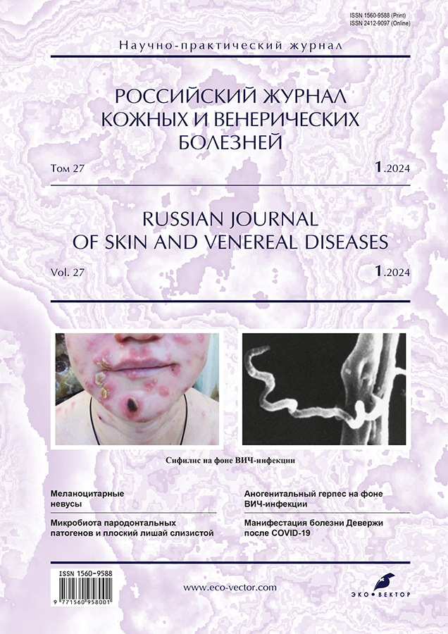
Photogallery. Anogenital herpes due to HIV infection

Cover Page


Cite item

Full Text

Open Access Open Access
Restricted Access Access granted
Restricted Access Subscription or Fee Access

Abstract

In patients with HIV infection, especially in the early stages and with preserved immunity, the same clinical manifestations of anogenital herpetic viral infection are observed as in immunocompetent individuals. As human immunodeficiency virus-associated (HIV) immunosuppression worsens in patients not taking antiretroviral therapy, the likelihood of developing atypical, disseminated forms of herpetic lesions increases. In severe immunodeficiency, the course of anogenital herpetic viral infection becomes more persistent, extensive, deep, long-term non-healing erosive and ulcerative rashes often develop, often spreading beyond their typical localization sites. In people living with human immunodeficiency virus with CD4+ T-lymphocyte counts below 50 cells/μl, erosions and ulcers caused by herpes simplex virus types 2 and/or 1 are often detected in the perianal area. In case of an atypical clinical picture, the diagnosis of anogenital herpetic viral infection is made based on the results of laboratory tests. The diagnostic standard is the detection of herpes simplex virus in scrapings from rashes using molecular genetic methods. In hypertrophic, tumor-like forms of the lesion, histological examination may be required.

All patients presented in the photo gallery are human immunodeficiency virus positive. He established the diagnosis of anogenital herpes based on the detection of one or simultaneously two types of herpes simplex virus in biomaterial from lesions using the polymerase chain reaction method. The explanations to the images indicate the stage and phase of human immunodeficiency virus infection in accordance with the Russian clinical classification, as well as the level of CD4+ T-lymphocytes at the time of diagnosis of anogenital herpetic viral infection.

Full Text

Restricted Access

About the authors

Sergey V. Prozherin

Sverdlovsk Regional Center for Prevention and Control of AIDS

Author for correspondence.
Email: progsherin@mail.ru
ORCID iD: 0000-0001-9956-4700
SPIN-code: 5354-4893
Scopus Author ID: 57221442199

dermatovenereologist

Russian Federation, Ekaterinburg

References

Supplementary files

Supplementary Files
Action
1. JATS XML
2. Fig. 1. Patient L., 29 years old. First clinical episode of genital herpes. In the biomaterial from erosion on the foreskin, herpes simplex virus type 2 was detected by polymerase chain reaction. On the inner layer of the foreskin, erosion with scalloped outlines against a background of erythema and single vesicles with serous contents are visible. Concomitant diagnosis: HIV infection (human immunodeficiency virus). Stage of secondary diseases (4A) is in the progression phase in the absence of antiretroviral therapy. The level of CD4+ T-lymphocytes is 308 cells/μl.

Download (727KB)
3. Fig. 2. Patient G., 29 years old. The first clinical episode of genital herpes caused by a combination of herpes simplex virus type 2 and herpes simplex virus type 1. Ulcerative-necrotic form. On the labia majora, perigenital area and perineal skin there are erosions and ulcers with areas of necrosis. Concomitant diagnosis: HIV infection. Stage of secondary diseases (4V) is in the progression phase in the absence of antiretroviral therapy. The number of CD4+ T-lymphocytes is 54 cells/μl.

Download (846KB)
4. Fig. 3. Patient Sh., 37 years old. First clinical episode of genital herpes caused by herpes simplex virus type 1 and herpes simplex virus type 2. Edema form. Against the background of pronounced edema and erythema of the left labia majora and adjacent skin areas, an irregularly shaped lesion is visible, partially covered with serous-hemorrhagic crusts. Concomitant diagnosis: HIV infection. Stage of secondary diseases (4A) in remission during antiretroviral therapy. The level of CD4+ T-lymphocytes is 576 cells/μl.

Download (820KB)
5. Fig. 4. Patient A., 41 years old. First clinical episode of genital herpes caused by herpes simplex virus type 2. Erosive-ulcerative form. Along the edge of the glans penis to the left of the frenulum there is an ulcer with a whitish coating on the surface and a rim of hyperemia along the periphery. Concomitant diagnosis: HIV infection. Stage of secondary diseases (4V) is in the progression phase in the absence of antiretroviral therapy. The absolute number of CD4+ T-lymphocytes is 40 cells/μl.

Download (535KB)
6. Fig. 5. Patient K., 45 years old. First clinical episode of genital herpes caused by herpes simplex virus type 2. Ulcerative-necrotic form. An extensive erosive and ulcerative lesion of irregular shape with clear boundaries, covering the skin of the penis, pubis and left inguinal-femoral-scrotal area. Along the periphery of the lesion, areas covered with gray-brown crusts are visible. Concomitant diagnosis: HIV infection. Stage of secondary diseases (4V) is in the progression phase in the absence of antiretroviral therapy. The absolute number of CD4+ T-lymphocytes is 2 cells/μl.

Download (586KB)
7. Fig. 6. Patient M., 36 years old. The first clinical episode of anogenital herpetic viral infection caused by herpes simplex virus type 2. Ulcer on the scrotum and erosive rashes on the inner surface of the thigh, inguinal-femoral-scrotal fold. Concomitant diagnosis: HIV infection. Stage of secondary diseases (4V) is in the progression phase in the absence of antiretroviral therapy. The level of CD4+ T-lymphocytes is 1 cell/μl.

Download (710KB)
8. Fig. 7. The same patient. Erosive and ulcerative lesions with serous-purulent plaque and a hyperemic rim along the periphery in the perianal region, intergluteal fold.

Download (557KB)
9. Fig. 8. Patient О., 32 years old. The first clinical episode of herpetic infection of the perianal skin. Erosive-ulcerative form. Erosive and ulcerative lesions in the perianal area. Using polymerase chain reaction, both types of herpes simplex virus were detected in scrapings from rashes. Concomitant diagnosis: HIV infection. Stage of secondary diseases (4V) is in the progression phase in the absence of antiretroviral therapy. The process manifested itself at a CD4+ T-lymphocyte level of 12 cells/μl.

Download (513KB)
10. Fig. 9. Patient L., 42 years old. The first clinical episode of herpetic infection of the perianal skin caused by herpes simplex virus type 2. Erosive-ulcerative form. Erosive and ulcerative lesions covered with fibrinous plaque in the perianal region, lower inner quadrant of the right buttock, intergluteal fold; scattered vesicles. Concomitant diagnosis: HIV infection. Stage of secondary diseases (4V) is in the progression phase in the absence of antiretroviral therapy. The level of CD4+ T-lymphocytes is 20 cell/μl.

Download (559KB)
11. Fig. 10. The same patient. The picture shows the condition after 2 months. Complete resolution of the rash occurred after 3 weeks of systemic administration of valacyclovir and antiretroviral therapy. In severe cases of anogenital herpesvirus infection, a standard course of systemic therapy with acyclic nucleosides does not always lead to resolution of clinical symptoms. In such cases, treatment is continued until complete regression.

Download (533KB)
12. Fig. 11. Patient A., 36 years old. Recurrent herpetic infection of the perianal skin. Erosions and ulcers with scalloped contours on the skin of the buttocks and intergluteal fold. Using the polymerase chain reaction method, herpes simplex virus type 2, herpes simplex virus type 1, cytomegalovirus and Epstein–Barr virus were detected in biological material from the rash. Concomitant diagnosis: HIV infection. Stage of secondary diseases (4V) is in the progression phase in the absence of antiretroviral therapy. The level of CD4+ T-lymphocytes is 4 cell/μl.

Download (611KB)
13. Fig. 12. The same patient. Extragenital localization of herpetic eruptions. Scrapings from the lesions revealed the same herpes viruses as in the perianal area. In severe immunodeficiency, herpes simplex can continuously recur, as was observed in this case.

Download (529KB)
14. Fig. 13. Patient B., 44 years old. Recurrence of genital herpes caused by herpes simplex virus type 2. Erosion on the inner layer of the foreskin. Concomitant diagnosis: HIV infection. Stage of secondary diseases (4A) in remission during antiretroviral therapy. The level of CD4+ T-lymphocytes is 452 cells/μl.

Download (557KB)
15. Fig. 14. Patient V., 44 years old. Recurrence of genital herpes caused by herpes simplex virus type 1 and herpes simplex virus type 2. Erosive elements and cracks on the frenulum and inner layer of the foreskin. Concomitant diagnosis: HIV infection. Stage of secondary diseases (4B) in remission during antiretroviral therapy. The level of CD4+ T-lymphocytes is 489 cells/μl.

Download (599KB)

Copyright (c) 2024 Eco-Vector


СМИ зарегистрировано Федеральной службой по надзору в сфере связи, информационных технологий и массовых коммуникаций (Роскомнадзор).
Регистрационный номер и дата принятия решения о регистрации СМИ: серия ПИ № ФС 77 - 86501 от 11.12.2023 г
СМИ зарегистрировано Федеральной службой по надзору в сфере связи, информационных технологий и массовых коммуникаций (Роскомнадзор).
Регистрационный номер и дата принятия решения о регистрации СМИ: серия ЭЛ № ФС 77 - 80653 от 15.03.2021 г
.


This website uses cookies

You consent to our cookies if you continue to use our website.

About Cookies