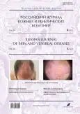
Photogallery. Juvenile hemangiomas

Cover Page


Cite item

Full Text

Open Access Open Access
Restricted Access Access granted
Restricted Access Subscription or Fee Access

Abstract

Juvenile hemangiomas can occur from the first months of life, with the prevalence of superficial juvenile hemangiomas (76.1%), more often solitary (83.3%).

Female infants predominate among the patients, with a 2:1 ratio.

Localization on the skin of the face and scalp is determined in 30% of cases; the remaining juvenile hemangiomas are located on the skin of the trunk and extremities.

Hemangiomas are mostly peripheral and small in size (10–20 mm), superficially and up to 5 mm deep.

The most frequent complication of juvenile hemangiomas is ulceration, both as a consequence of exophytic growth and treatment methods.

The predominance of arterial intramedullary blood flow was found at ultrasound diagnosis, however, variants of 2-phase blood flow can also occur, which requires additional ultrasound examination of all cases of juvenile hemangiomas.

We offer the publication of a photo gallery on this problem.

Full Text

Restricted Access

About the authors

Vladislav V. Dubensky

Tver State Medical University

Author for correspondence.
Email: dubensky.vladislav@yandex.ru
Russian Federation, Tver

Valeriy V. Dubenskiy

Tver State Medical University

Email: valerydubensky@yandex.ru

MD, Dr. Sci. (Med.), Professor

Russian Federation, Tver

References

Supplementary files

Supplementary Files
Action
1. JATS XML
2. Fig. 1. Patient C., 1.5 months old. Diagnosis: "Focal superficial juvenile hemangioma". The process is located on the skin of the chest, bright red, irregular outlines, 4×8 cm in size, elevated above the skin level, flat in shape, there are separate sunken areas on the surface (with decreasing infiltrate). On the periphery, the skin is white with individual capillary hemangiomas 1 to 2 mm in size.

Download (624KB)
3. Fig. 2. Patient C., 8 months old. Diagnosis: "Focal superficial hemangioma of the breast skin (telangiectatic variant)". There is an area of affected skin 4×7 cm with clear boundaries, represented by telangiectasias with insignificant lumen of healthy skin.

Download (493KB)
4. Fig. 3. Patient B., 6 months old. Diagnosis: "Combined juvenile hemangioma of the scalp": а ― the neoplasm is located in the frontal and temporal region, represented by a vascular node, red-violet in color, up to 5 cm in diameter, with clear boundaries, 1.5–2 cm above the skin level; there is an area of fibrosis in the center of the hemangioma; b (ultrasound examination) ― sonogram of combined juvenile hemangioma: Biphasic (arterial) blood flow type is detected.

Download (777KB)
5. Fig. 4. Patient D., 4 months old. Diagnosis: "Combined juvenile hemangioma of the left cheek skin". The process is represented by a cherry-red tumor-like neoplasm, up to 12 mm in diameter, up to 1 cm above the skin level. It is located against the background of visibly unchanged skin.

Download (524KB)
6. Fig. 5. Same patient. Color Doppler mapping of a combined juvenile hemangioma: its homogeneous echostructure revealed (а); pulsed-wave Doppler imaging of combined juvenile hemangioma: monophasic blood flow type established (b).

Download (586KB)
7. Fig. 6. Patient N., 11 months old. Diagnosis: "Segmental capillary angiodysplasia of the right side of the face ("Wine spot")". The process is located on the skin of the right side of the face, spreads to the temporal, cheek and periorbital areas, presented with pink-red erythema, with cyanotic component, clear borders, not elevated above the skin level. The skin is pale during vitropresis. It is necessary to differentiate with the superficial form of hemangioma.

Download (720KB)
8. Fig. 7. Patient N., 8 months old. Diagnosis: "Superficial juvenile hemangioma of the right labia with a laceration". The process has spread to the skin of the pubis, labia majora, perianal area; the elements are pink-red in color with single luminescence. There is asymmetry due to the right labia (due to inflammation and subsequent lymphostasis). In the center of the lip there was an ulcerous defect measuring 8×3 mm, of irregular outline, covered with fibrinous film. The defect was a consequence of local chemodestruction.

Download (361KB)
9. Fig. 8. Patient Z., 4 months old. Diagnosis: "Combined hemangioma of the skin of the chin area with ulceration". The tumor-like mass covers the entire chin, red-blueish in color with a linear ulcerous defect formed as a result of exophytic hemangioma growth.

Download (407KB)
10. Fig. 9. Patient K., 1 month old. Diagnosis: "Superficial juvenile hemangioma of the anterior axillary line skin". The process is represented by a red capillary hemangioma with indistinct boundaries, spreading from the skin of the chest to the right axillary fold. The borders are clear; the outlines are irregular, 5×8 cm in size.

Download (260KB)
11. Fig. 10. The same patient. а ― After 1 month of treatment with topical beta-adrenoblocker; the site of the resolved hemangioma had a lividity-like rash; b ― after 3 months of treatment with a topical beta-adrenoblocker.

Download (1MB)

Copyright (c) 2023 Eco-Vector

License URL: https://eco-vector.com/for_authors.php#07

СМИ зарегистрировано Федеральной службой по надзору в сфере связи, информационных технологий и массовых коммуникаций (Роскомнадзор).
Регистрационный номер и дата принятия решения о регистрации СМИ: серия ПИ № ФС 77 - 86501 от 11.12.2023 г
СМИ зарегистрировано Федеральной службой по надзору в сфере связи, информационных технологий и массовых коммуникаций (Роскомнадзор).
Регистрационный номер и дата принятия решения о регистрации СМИ: серия ЭЛ № ФС 77 - 80653 от 15.03.2021 г
.


This website uses cookies

You consent to our cookies if you continue to use our website.

About Cookies