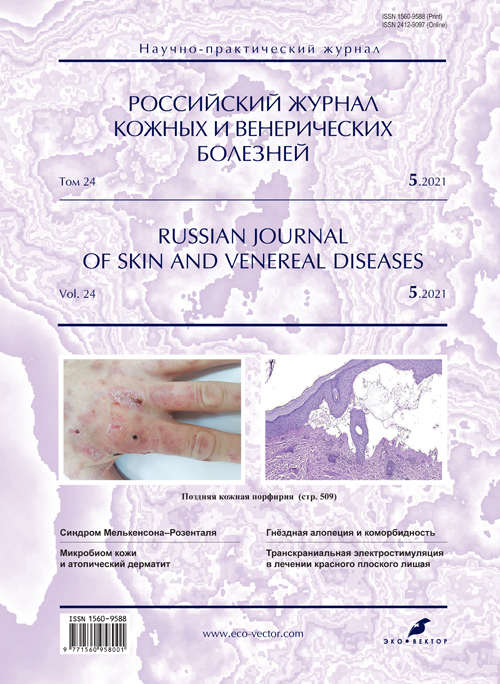
Photo gallery. Superficial mycoses of the skin
- 作者: Yakovlev A.B.1, Maximov I.S.1
-
隶属关系:
- I.M. Sechenov First Moscow State Medical University (Sechenov University)
- 期: 卷 24, 编号 5 (2021)
- 页面: 525-528
- 栏目: PHOTO GALLERY
- ##submission.dateSubmitted##: 18.01.2022
- ##submission.dateAccepted##: 14.02.2022
- ##submission.datePublished##: 12.09.2021
- URL: https://rjsvd.com/1560-9588/article/view/96704
- DOI: https://doi.org/10.17816/dv96704
- ID: 96704
如何引用文章
全文:
详细
Superficial fungal infections of the skin are common on all continents. Errors in the diagnosis and treatment of these patients are often observed. The lack of symmetry of the process, erythematous-squamous foci with a raised roller and more pronounced peeling along the periphery, polycyclic and annular outlines, as well as the presence of papules and pustules in the lesion can give a hint in making a correct diagnosis. Laboratory tests (microscopy and seeding) are mandatory for accurate diagnosis.
This article presents photos with anamnesis of the disease and treatment options for common superficial fungal infections of the skin.
全文:
Пациент, 16 лет, болен около 5,5 мес, когда появились высыпания на коже плеч, сопровождавшиеся зудом (рис. 1). Проживает в частном доме с дедушкой и бабушкой. Имеются домашние животные, две козы, кролики. Ногтевые пластинки стоп и кистей не поражены. Обращался к дерматологу: взят соскоб на микроскопию и выполнен посев.
Рис. 1. Микоз гладкой кожи. / Fig. 1. Mycosis of smooth skin.
Получен рост гриба Trichophyton rubrum. В течение 2 мес проводилось лечение только наружными препаратами (тербинафин крем и повидон-йод) с хорошим эффектом. По завершении лечения мицелий патогенных грибов в соскобе троекратно не обнаружен, однако через 3–4 нед кожный процесс возоб- новился.
Пациент проходит обследование с целью назначения комбинированной терапии.
Пациент, 55 лет, болен 17 дней; обратился к дерматологу спустя 2 нед от появления высыпаний (рис. 2). Самостоятельно никакие препараты не применял. В течение последних двух дней беспокоит зуд. В соскобе обнаружен мицелий гриба. Установлен диагноз микоза кожи, онихомикоз стоп. Применяет крем тербинафин по назначению врача.
Рис. 2. Микоз кожи правой голени. / Fig. 2. Mycosis of the skin of the right leg.
Пациент, 48 лет, болен около 1,5 мес, когда стали появляться высыпания с умеренным зудом на коже груди; кроме того, имеются проявления микоза кожи и ногтевых пластинок стоп (рис. 3).
Рис. 3. Микоз гладкой кожи груди, симулирующий склеродермию. / Fig. 3. Mycosis of the smooth skin of the breast, simulating scleroderma.
По настоянию матери обратился в кожно-венерологический диспансер, где после обследования выставлен диагноз: «Микоз кожи и ногтевых пластинок стоп, микоз гладкой кожи». Назначены повидон-йод, нафтифин крем, а также обследование для назначения комбинированной терапии.
Пациент, 70 лет, болен 12 дней, когда случайно заметил очаг на коже правой голени (рис. 4). Пенсионер. Проживает постоянно в Москве, но в период с мая по октябрь находится в деревне в Тамбовской области. Имеет двух собак и двух кошек.
Рис. 4. Микроспория гладкой кожи. / Fig. 4. Microsporia of smooth skin.
В соскобе обнаружен мицелий патогенного гриба с поражением волос по смешанному типу ecto- и endothrix. В посеве получен рост Microsporum audouinii.
Назначено лечение: наружно на область высыпаний повидон-йод 2 раза в день с последующим нанесением крема сертаконазола, курс 12 дней.
Пациент выполнял назначение частично: смазывал очаг повидон-йодом и дегтярной мазью по «народному» рецепту. По завершении лечения в соскобах, выполненных дважды, мицелий не обнаружен.
Пациентка Ж., 28 лет; высыпания появились после контакта с бездомной кошкой в доме отдыха. На фото отчётливо видно зеленоватое свечение в лучах лампы Вуда (рис. 5). При осмотре наблюдаются множественные очаги на коже туловища и нижних конечностей. Поражений волосистой части головы не обнаружено.
Рис. 5. Микроспория гладкой кожи: свечение в лучах лампы Вуда. / Fig. 5. Microsporia of smooth skin: glow in the rays of a Woods lamp.
Назначена системная и местная терапия тербинафином.
Пациентка, 47 лет, обнаружен мицелий патогенных грибов (рис. 6). Назначен раствор Фукорцин 1 раз в день, 7 дней; сертаконазол крем 2 раза в день. Кожный проц3есс разрешился.
Рис. 6. Микроспория гладкой кожи, симулирующая стрептодермию. / Fig. 6. Microsporia of smooth skin simulating streptoderma.
Сразу после лечения в трёх соскобах с интервалом 5 дней мицелий патогенных грибов не обнаружен; в четвёртом анализе через 20 дней ― не обнаружен.
Пациентка Г., 54 года; высыпания субмаммарных складок беспокоят в течение 2 нед (рис. 7). Из анамнеза известно о наличии у пациентки метаболического синдрома. На консультации выполнен соскоб кожи, биоматериал отправлен в лабораторию на микроскопию и культуральное исследование. При микроскопии обнаружен псевдомицелий, в посеве ― Candida spp.
Рис. 7. Кандидоз субмаммарной складки. / Fig. 7. Submammary fold candidiasis.
Рекомендовано: флуконазол в дозе 150 мг в первый день, затем по 50 мг/сут ежедневно в течение 3 нед; наружно клотримазол крем 2 раза/сут. Консультация эндокринолога обязательна.
Пациент, 58 лет, болен около 3 нед, когда появились высыпания на коже правой подмышечной области (рис. 8). Лечился самостоятельно детским кремом. Периодически беспокоит зуд. Пользовался услугами частнопрактикующего врача: анализ на грибы не выполнен; назначен крем вида клотримазол/гентамицин/бетаметазон с хорошим, но нестойким эффектом.
Рис. 8. Микоз кожи крупной складки в аксиллярной области справа. / Fig. 8. Mycosis of the skin of a large fold in the axillary region on the right.
Пациентка, 59 лет; сопутствующее заболевание: нарушение толерантности к глюкозе (рис. 9).
Рис. 9. Кандидоз мелких складок (межпальцевая складка 3–4-го пальцев левой кисти). / Fig. 9. Candidiasis of small folds (interdigital fold of 3–4 fingers of the left hand).
Назначения: водный режим, по возможности низкоуглеводная диета; наружно: утром ― Фукорцин 1 раз в день, 8 дней; вечером ― первые 5 дней лечения крем вида клотримазол/бетаметазон, затем в течение 14 дней эконазол крем 1 раз в день.
Пациентка, 32 года, больна в течение 4 мес. Проживает в селе, обращалась к местному фельдшеру: диагноз озвучен не был; назначались сначала мази с антибиотиками (без эффекта), затем серно-салициловая мазь, каждый раз в комбинации с 2% йодной настойкой.
Всего у пациентки на голове насчитано 15 мелких очагов (до 1 см) и один крупный (5 см) (рис. 10). Назначено комбинированное лечение с тербинафином внутрь.
Рис. 10. Инфильтративно-нагноительная трихофития волосистой части головы, нагноительная фаза. / Fig. 10. Infiltrative suppurative trichophytosis of the scalp, suppurative phase.
Пациент Г., 63 года, обратился на консультацию с поражением кожи в области левого коленного сустава (рис. 11). При осмотре наблюдаются также поражения стоп и ногтей. При микроскопии соскобов чешуек кожи и ногтей обнаружен мицелий патогенных грибов, в посеве ― рост Trichophyton rubrum.
Рис. 11. Рубромикоз гладкой кожи в области разгибательной поверхности левого коленного сустава. / Fig. 11. Rubromycosis of smooth skin in the area of the extensor surface of the left knee joint.
По результатам общего анализа и биохимии крови назначена комбинированная терапия: утром per os тербинафин в дозе 250 мг/сут после еды под контролем биохимического анализа крови 1 раз в 4 нед; наружно тербинафин крем 2 раза/сут. Повторная консультация ― через 1 мес.
Пациент К., 48 лет; кроме поражения кожи нижних конечностей наблюдаются высыпания на коже спины и верхних конечностей (рис. 12). Из анамнеза известно, что пациент неоднократно обращался к врачам: под вопросом выставлялся диагноз аллергической реакции; применял крем вида клотримазол/гентамицин/бетаметазон в течение 3 нед без терапевтического эффекта. В дальнейшем другим специалистом выполнена микроскопия гнойного отделяемого и чешуек кожи: обнаружен мицелий патогенных грибов. Рекомендована системная терапия: тербинафин в дозе 250 мг/сут в течение 3 нед без применения местных препаратов.
Рис. 12. Микоз гладкой кожи в области сгибательной поверхности коленного сустава: после 3 нед монолечения системным препаратом. / Fig. 12. Mycosis of smooth skin in the area of the flexor surface of the knee joint: after 3 weeks of monotherapy with a systemic drug.
На консультации в клинике кожных и венерических болезней имени В.А. Рахманова пациенту рекомендован итраконазол в дозе 200 мг/сут в течение 4 нед, наружно сертаконазол крем 2 раза/сут. При повторной консультации через 4 нед процесс регрессировал на 90%. Рекомендовано продолжить местную терапию.
作者简介
Alexey Yakovlev
I.M. Sechenov First Moscow State Medical University (Sechenov University)
编辑信件的主要联系方式.
Email: aby@rinet.ru
ORCID iD: 0000-0001-7073-9511
MD, PhD, Docent
俄罗斯联邦, MoscowIvan Maximov
I.M. Sechenov First Moscow State Medical University (Sechenov University)
Email: maximov.is@mail.ru
ORCID iD: 0000-0003-2850-2910
MD, Assistant
俄罗斯联邦, Moscow参考
补充文件


