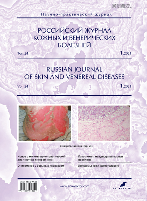
Skin lymphomas (photogallery)
- 作者: Olisova O.Y.1, Grekova E.V.1, Chernova N.G.2
-
隶属关系:
- I.M. Sechenov First Moscow State Medical University (Sechenov University)
- National Research Center for Hematology
- 期: 卷 24, 编号 1 (2021)
- 页面: 101-106
- 栏目: PHOTO GALLERY
- ##submission.dateSubmitted##: 07.07.2021
- ##submission.dateAccepted##: 07.07.2021
- ##submission.datePublished##: 15.02.2021
- URL: https://rjsvd.com/1560-9588/article/view/75794
- DOI: https://doi.org/10.17816/dv75794
- ID: 75794
如何引用文章
全文:
详细
Skin lymphomas are a heterogeneous group of lymphoproliferative diseases characterized by clonal proliferation and primary accumulation of tumor lymphocytes in the skin with the possibility of secondary extracutaneous spread (lymph nodes, blood, spleen, lungs, liver). 65% of all skin lymphomas originate from mature T-cells, 25% from mature B-cells, 10% originate from natural killer cells (NK).
全文:
WHO-EORTC-КЛАССИФИКАЦИЯ ПЕРВИЧНЫХ ЛИМФОМ КОЖИ (ПЕРЕСМОТР 2018 ГОДА)
Т-клеточные лимфомы кожи
1.Грибовидный микоз
1.1. Фолликулотропная форма
1.2. Педжетоидный ретикулёз
1.3. Синдром гранулематозной вялой кожи
- Синдром Сезари
- Т-клеточная лейкемия/лимфома взрослых
- Первичные кожные CD30+ лимфопролиферативные заболевания
4.1. Первичная кожная анапластическая крупноклеточная лимфома
4.2. Лимфоматоидный папулёз
- Панникулитоподобная Т-клеточная лимфома подкожной жировой клетчатки
- Экстранодальная NK/T-клеточная лимфома,
назальный тип - Хроническая активная EBV-инфекция
- Первичные кожные периферические Т-клеточные лимфомы, редкие варианты
8.1. Первичная кожная γ/δ Т-клеточная лимфома
8.2. Первичная кожная агрессивная эпидермотропная CD8+ Т-клеточная лимфома
8.3. Т-клеточное CD4+ лимфопролиферативное заболевание из лимфоцитов малого и среднего размеров
8.4. Первичная кожная акральная CD8+ Т-клеточная лимфома
- Первичная кожная периферическая Т-клеточная лимфома, неуточнённая.
В-клеточные лимфомы кожи
- Первичная кожная В-клеточная лимфома маргинальной зоны
- Первичная кожная лимфома из клеток фолликулярного центра
- Первичная кожная диффузная крупноклеточная В-клеточная лимфома, тип нижних конечностей
- EBV+ слизисто-кожная язва
- Внутрисосудистая крупноклеточная В-клеточная лимфома
В группе больных Т-клеточной лимфомой кожи подавляющее большинство (75–80%) составляют больные грибовидным микозом.
Рис. 1. Пятнистая стадия классической формы грибовидного микоза. / Fig. 1. Spotted stage of the classical form of fungal mycosis
Рис. 2. Бляшечная стадия классической формы грибовидного микоза. / Fig. 2. Plaque stage of the classical form of fungal mycosis
Рис. 3. Лимфоматоидный папулёз, тип С, ассоциированный с грибовидным микозом. / Fig. 3. Lymphatic papulosis, type C, associated with fungal mycosis
Рис. 4. Панникулитоподобная Т-клеточная лимфома. / Fig. 4. Panniculitis-like T-cell lymphoma
Рис. 5. Склеродермоподобная форма грибовидного микоза. / Fig. 5. Scleroderm-like form of fungal mycosis.
Рис. 6. Сиринготропная форма грибовидного микоза. Фото Коломейцева О.А. / Fig. 6. Syringotropic form of fungal mycosis. Photo by O. Kolomeytsev.
Рис. 7. Грибовидный микоз: пятна и бляшки. / Fig. 7. Fungal mycosis: spots and plaques.
Рис. 8. Фолликулотропный подтип грибовидного микоза, ассоциированный с фолликулярным муцинозом. / Fig. 8. Folliculotropic subtype of fungal mycosis associated with follicular mucinosis.
Рис. 9. Опухолевая стадия грибовидного микоза: множественные узлы. / Fig. 9. Tumor stage of fungal mycosis: multiple nodes.
Рис. 10. Опухолевая стадия грибовидного микоза: узлы на лице. / Fig. 10. Tumor stage of fungal mycosis: nodes on the face.
Рис. 11. Первичная кожная анапластическая крупноклеточная лимфома: рупеоидные высыпания кожи спины. Фото Черновой Н.Г. / Fig. 11. Primary cutaneous anaplastic large cell lymphoma: rupeoid eruptions of the back skin. Photo by N.G. Chernovaya.
Рис. 12. Первичная кожная CD4+ плеоморфная Т-клеточная лимфома: а ― пятна и бляшки на коже туловища; б ― узел на стопе. / Fig. 12. Primary cutaneous CD4+ pleomorphic T-cell lymphoma: а ― spots and plaques on the skin of the trunk; б ― knot on the foot.
Рис. 13. Грибовидный микоз: множественные узлы на голове. / Fig. 13. Fungal mycosis: multiple nodes on the head.
Рис. 14. Синдром гранулематозной вялой кожи. / Fig. 14. Syndrome of granulomatous flaccid skin.
Рис. 15. Пойкилодермическая форма грибовидного микоза. / Fig. 15. Poikilodermic form of fungal mycosis.
作者简介
Olga Olisova
I.M. Sechenov First Moscow State Medical University (Sechenov University)
编辑信件的主要联系方式.
Email: olisovaolga@mail.ru
ORCID iD: 0000-0003-2482-1754
SPIN 代码: 2500-7989
Dr. Sci. (Med.), Professor
俄罗斯联邦, MoscowEkaterina Grekova
I.M. Sechenov First Moscow State Medical University (Sechenov University)
Email: grekova_kate@mail.ru
ORCID iD: 0000-0002-7968-9829
SPIN 代码: 8028-5545
Assistant Professor
俄罗斯联邦, MoscowNatalya Chernova
National Research Center for Hematology
Email: grekova_kate@mail.ru
俄罗斯联邦, Moscow
参考
补充文件


