


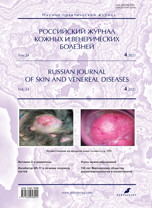
Множественная цилиндрома кожи головы
- Авторы: Вертиева Е.Ю.1, Теплякова К.С.1
-
Учреждения:
- Первый Московский государственный медицинский университет имени И.М. Сеченова (Сеченовский Университет)
- Выпуск: Том 24, № 4 (2021)
- Страницы: 329-334
- Раздел: ДЕРМАТООНКОЛОГИЯ
- Статья получена: 16.09.2021
- Статья одобрена: 02.12.2021
- Статья опубликована: 15.07.2021
- URL: https://rjsvd.com/1560-9588/article/view/80212
- DOI: https://doi.org/10.17816/dv80212
- ID: 80212

Цитировать
Полный текст

Аннотация
Цилиндрома ― редкое доброкачественное эпителиальное новообразование из придатков кожи, типичной локализацией которого являются голова и шея. Патогенез заболевания неизвестен, однако сегодня принято считать, что цилиндромы развиваются из эпителиальных протоков эккринных или апокринных желёз, либо представляют собой неопластическую пролиферацию эпителиальных стволовых клеток.
Клинически цилиндрома представлена единичными или множественными гладкими отграниченными от здоровой кожи узелками и узлами бледно-розового цвета, на поверхности которых часто видны ветвящиеся сосуды.
Выделяют спорадическую, генетически не обусловленную цилиндрому, и множественную, наследуемую по аутосомно-доминантному типу.
При множественных цилиндромах из-за слияния и увеличения размеров узелков и узлов на коже волосистой части головы образуется поражение, внешне напоминающее тюрбан, поэтому иногда цилиндрому называют «тюрбанной опухолью».
Лечение множественной цилиндромы представляет серьёзную междисциплинарную проблему в связи с большой площадью поражения, хорошим кровоснабжением волосистой части головы и склонностью заболевания к рецидивированию. Описанные в литературе методы лечения включают иссечение традиционным хирургическим методом, электрокоагуляцию, лазер, микрографическую хирургию по Мосу.
В статье приводится редкий клинический случай развития множественной цилиндромы у молодого мужчины.
Ключевые слова
Полный текст
ВВЕДЕНИЕ
Цилиндрома ― редкая доброкачественная эпителиальная опухоль из придатков кожи, происхождение которой по-прежнему остаётся спорным.
Заболевание манифестирует на втором или третьем десятке жизни; женщины заболевают в два раза чаще мужчин [1]. Выделяют спорадические цилиндромы и множественные: последние наследуются по аутосомно-доминантному типу, могут являться составной частью кожного синдрома CYLD, ранее описанного в литературе как синдром Брука–Шпиглера. В случае одиночного поражения связь с семейным анамнезом отсутствует [2].
Патогенез заболевания остаётся неизвестным. Ранее на основании сходства клеток цилиндромы с клетками эккринных потовых желёз придерживались эккринной теории происхождения опухоли [3]. Сегодня принято считать, что цилиндромы развиваются из эпителиальных протоков эккринных или апокринных желёз, либо представляют собой неопластическую пролиферацию эпителиальных стволовых клеток [4]. В литературе описана также теория происхождения цилиндром из опухоли волосяного фолликула [5].
Цилиндрома представляет собой отграниченную от эпидермиса опухоль, состоящую из цилиндрических и овальных гнёзд базальных клеток в дерме, сформированных в виде мозаики. Клетки центральной части опухоли в отличие от клеток периферической части окрашены менее ярко. Опухоль окружена гиалиновой мембраной, состоящей из белков внеклеточного матрикса (включая коллаген IV и VII и ламинин-332) в базальной мембране кожи [2, 6].
Типичная клиническая картина опухоли представлена единичными или множественными гладкими, отграниченными от здоровой кожи узелками и узлами бледно-розового цвета, на поверхности которых часто видны ветвящиеся сосуды [6]. Характерной локализацией являются голова и шея, однако в литературе описаны и более редкие локализации: слуховой проход, орбитальная область, конечности, туловище [2, 7, 8].
Цилиндрома имеет выраженную тенденцию к росту и слиянию, при этом образуется характерный вид опухолевых разрастаний, так называемый «тюрбан». Несмотря на то, что цилиндрома относится к доброкачественным образованиям, в редких случаях возможна малигнизация. К клиническим признакам, указывающим на злокачественную трансформацию, относят быстрый рост образования, болезненность, изменение цвета и кровоточивость [1].
Лечение множественной цилиндромы представляет собой серьёзную междисциплинарную проблему в связи с большой площадью поражения и хорошим кровоснабжением кожи волосистой части головы.
Описаны различные методы терапии данного заболевания: иссечение традиционным хирургическим методом, электрокоагуляция, лазер, микрографическая хирургия по Мосу. При выборе метода следует учитывать распространённость поражения, локализацию, склонность заболевания к рецидивированию.
При обширном поражении кожного покрова и множественных сливающихся узлах удаление производят в несколько этапов. Следует помнить, что цилиндромы склонны к рецидивированию. В случаях рецидива предпочтение следует отдать методу микрографической хирургии по Мосу [6].
Данное заболевание требует дифференциальной диагностики с нейрофиброматозом I типа (болезнь Реклингхаузена). В отличие от цилиндромы опухолевые образования при нейрофиброматозе (кожные и подкожные нейрофибромы) имеют цвет нормальной кожи, розовато-сиреневый или коричневый, чаще всего бывают мягкой эластичной консистенции. Над подкожными нейрофибромами имеется грыжевое выпячивание, легко поддающееся вдавлению при пальпации [9].
Представляем клинический случай множественной цилиндромы волосистой части головы.
ОПИСАНИЕ КЛИНИЧЕСКОГО СЛУЧАЯ
Больной М., 27 лет, обратился в лечебно-диагностическое отделение кожных и венерических болезней Первого МГМУ им. И.М. Сеченова с жалобами на множественные образования волосистой части головы без субъективных ощущений.
Анамнез заболевания. Считает себя больным около 9 лет, когда впервые заметил появление небольшого узелка на коже волосистой части головы. Наблюдался по месту жительства с диагнозом множественной цилиндромы волосистой части головы, где проводилась периодическая электрокоагуляция новообразований с временным положительным эффектом.
Сопутствующая патология. Код по МКБ-10: F71 Умственная отсталость умеренная.
Употребление алкоголя, курение отрицает, аллергические реакции отрицает. Наследственность не отягощена.
Результаты обследования
Объективно. На момент осмотра процесс локализуется на коже волосистой части головы. Поражение кожи носит хронический подостровоспалительный характер. Высыпания представлены множеством мономорфных узелков и узлов размером от 5 мм до 5 см, полушаровидной формы с гладкой поверхностью, округлых очертаний, чётко отграниченных от окружающей кожи. Цвет образований от бледно-розового до красноватого, консистенция плотная. Субъективные ощущения отсутствуют (рис. 1).

Рис. 1. Больной М., 27 лет. Клинико-морфологическая картина множественной цилиндромы: а ― на коже волосистой части головы узел плотной консистенции размером 5 см, полушаровидной формы, с гладкой поверхностью и чёткими границами, бледно-розового цвета; на поверхности видны ветвящиеся сосуды; б, в ― на коже волосистой части головы множество мономорфных узелков и узлов размером от 5 мм до 3 см, плотной консистенции, полушаровидной формы, с гладкой поверхностью, чёткими границами, от бледно-розового до красноватого цвета. / Fig. 1. Patient M., 27 years old. The clinical and morphological presentations of multiple cylindroma: а ― on the skin of the scalp, a node of dense consistency with a size of 5 cm, hemispherical shape, with a smooth surface and clear boundaries, pale pink; branching vessels are visible on the surface; б, в ― on the skin of the scalp, many monomorphic nodules and nodes ranging in size from 5 mm to 3 cm, dense consistency, hemispherical shape, with a smooth surface, clear boundaries, ranging in color from pale pink to reddish.
Дерматоскопическое исследование: неоангиогенез, ветвящиеся сосуды. Картина, характерная для диагноза множественной цилиндромы кожи головы (рис. 2).

Рис. 2. Больной М., 27 лет. Дерматоскопическая картина: неоангиогенез, ветвящиеся сосуды. Картина, характерная для диагноза множественной цилиндромы кожи головы. / Fig. 2. Patient M., 27 years old. Dermatoscopic features: neoangiogenesis, branching vessels. The dermoscopic pattern is typical for the diagnosis of multiple scalp cylindroma.
Гистологическое исследование: опухоль локализована в дерме и подкожной жировой клетчатке, состоит из комплексов, имеющих вид долек различного размера, овальной или цилиндрической формы, чётко отграниченных друг от друга, окружённых толстыми эозинофильными гиалиноподобными отложениями и фиброзной тканью.
Диагноз. На основании жалоб, клинической картины и проведённых исследований был выставлен диагноз: «Множественная цилиндрома кожи головы».
Для определения тактики лечения необходимо привлечение смежных специалистов, так как объём поражения не позволяет проводить лечение в условиях клиники кожных и венерических болезней им. В.А. Рахманова.
Пациент направлен на консультацию к хирургу для обсуждения объёма операционного вмешательства. Запланирована консультация клинического генетика.
ОБСУЖДЕНИЕ
Приведённый клинический случай представляет интерес в связи с несколькими аспектами.
По данным литературы, множественная цилиндрома имеет генетическую основу. Однако в описанном случае генетическая наследственность не выявлена из-за отсутствия генетического тестирования и отсутствия семейного анамнеза цилиндромы. В связи с выявленной у пациента умственной отсталостью можно предположить наличие генетического дефекта, однако невозможно точно сказать о наличии генетической основы заболевания.
Клинические проявления заболевания у пациента соответствуют литературным данным: начало заболевания в подростковом периоде, локализация на волосистой части головы, поражение хронического подостровоспалительного характера, склонное к рецидивированию. Высыпания представлены множеством мономорфных узелков и узлов размером от 5 мм до 5 см, полушаровидной формы с гладкой поверхностью, округлых очертаний, чётко отграниченных от окружающей кожи. Цвет образований от бледно-розового до красноватого, консистенция плотная. Субъективные ощущения отсутствуют.
Цилиндрома ― медленно растущая доброкачественная опухоль. «Тюрбанная опухоль» может достигать огромных размеров и приводить к обезображивающему дефекту.
Для постановки диагноза используются следующие критерии:
- манифестация в подростковом возрасте;
- наличие единичных или множественных опухолей у родственников;
- гистологически верифицированные цилиндромы, трихоэпителиомы, спираденомы;
- мутация в гене CYLD на хромосоме 16q12-q13;
- образования опухолей сальных и паращитовидных желёз.
При синдроме Брука–Шпиглера, а также множественных цилиндромах возможна малигнизация опухоли. Признаками озлокачествления являются быстрый рост, изъязвление, красно-фиолетовая окраска, иногда болезненность. Для исключения злокачественности цилиндромы используют магнитно-резонансную и компьютерную томографию. При этом разрушение костной ткани на снимках является признаком злокачественной трансформации очага.
Метастазирование цилиндромы происходит крайне редко и осуществляется в лимфатические узлы шеи, подмышечные и лимфатические узлы средостения.
Для лечения применяют деструктивные методы: хирургию по Мосу, лазерную абляцию, электрокоагуляцию и радиоволновую деструкцию. При неоперабельных опухолях возможно назначения лучевой терапии. Однако основным методом лечения по-прежнему остаётся широкое иссечение опухоли в связи с её частым рецидивированием с последующей пластикой свободным лоскутом.
Таким образом, все пациенты с синдромом Брука или множественными цилиндромами должны регулярно проходить обследование у онкодерматолога; выполнять магнитно-резонансную и компьютерную томографию для исключения трансформации в цилиндрокарциному.
ЗАКЛЮЧЕНИЕ
Исходя из нашего опыта, ранняя диагностика и свое-временное удаление новых образований при рецидивировании являются ключевыми моментами в ведении пациентов с диагнозом множественной цилиндромы. Это позволяет избежать обширного хирургического вмешательства и достичь эффекта при минимальной травматизации. Все пациенты с установленным диагнозом должны регулярно проходить профилактический осмотр у онкодерматолога для исключения малигнизации образований.
ДОПОЛНИТЕЛЬНО
Источник финансирования. Работа выполнена по инициативе авторов без привлечения финансирования.
Конфликт интересов. Авторы декларируют отсутствие явных и потенциальных конфликтов интересов, связанных с содержанием настоящей статьи.
Вклад авторов. Е.Ю. Вертиева ― получение, анализ и интерпретация результатов, внесение существенных правок с целью повышения научной ценности статьи; К.С. Теплякова ― концепция и дизайн исследования, написание рукописи. Все авторы подтверждают соответствие своего авторства международным критериям ICMJE (все авторы внесли существенный вклад в разработку концепции, проведение исследования и подготовку статьи, прочли и одобрили финальную версию перед публикацией).
Согласие пациента. Пациент добровольно подписал информированное согласие на публикацию персональной медицинской информации в обезличенной форме.
ADDITIONAL INFO
Funding source. This work was not supported by any external sources of funding.
Competing interests. The authors declare that they have no competing interests.
Author contribution. E.Y. Vertieva ― obtaining, analyzing and interpreting the results, editing to increase the scientific value of the article. K.S. Teplyakova ― the concept the design of the study, drafting the manuscript. The authors made a substantial contribution to the conception of the work, acquisition, analysis of literature, drafting and revising the work, the final approval of the version to be published and agree to be accountable for all aspects of the work.
Patients permission. The patient voluntarily signed an informed consent to the publication of personal medical information in an impersonal form.
Об авторах
Екатерина Юрьевна Вертиева
Первый Московский государственный медицинский университет имени И.М. Сеченова (Сеченовский Университет)
Автор, ответственный за переписку.
Email: ivertieva@gmail.com
ORCID iD: 0000-0002-1088-2911
SPIN-код: 3712-8453
к.м.н.
Россия, МоскваКсения Сергеевна Теплякова
Первый Московский государственный медицинский университет имени И.М. Сеченова (Сеченовский Университет)
Email: hvpvea@gmail.com
ORCID iD: 0000-0002-0428-5627
SPIN-код: 5983-9344
аспирант
Россия, МоскваСписок литературы
- Kuklani R.M., Glavin F.L., Bhattacharyya I. Malignant cylindroma of the scalp arising in a setting of multiple cylindromatosis: a case report // Head Neck Pathol. 2009. Vol. 3, N 4. Р. 315–319. doi: 10.1007/s12105-009-0138-x
- Chauhan D.S., Guruprasad Y. Dermal cylindroma of the scalp // Natl J Maxillofac Surg. 2012. Vol. 3, N 1. Р. 59–61. doi: 10.4103/0975-5950.102163
- Munger B.L., Graham J.H., Helwig E.B. Ultrastructure and histochemical characteristics of dermal eccrine cylindroma (turban tumor) // J Invest Dermatol. 1962. Vol. 39. Р. 577–595.
- Tunggal L., Ravaux J., PeschM., at al. Defective laminin 5 processing in cylindroma cells // Am J Pathol. 2002. Vol. 160, N 2. Р. 459–468. doi: 10.1016/S0002-9440(10)64865-1
- Massoumi R., Podda M., Fässler R., Paus R. Cylindroma as tumor of hair follicle origin // J Invest Dermatol. 2006. Vol. 126, N 5. Р. 1182–1184. doi: 10.1038/sj.jid.5700218
- Adam M.P., Ardinger H.H., Pagon R.A., et al., editors. Gene Reviews [интернет]. Seattle (WA): University of Washington, Seattle; 1993–2021. Режим доступа: https://www.ncbi.nlm.nih.gov/books/NBK1116/. Дата обращения: 15.11.2021.
- Schwarz D., Drebber U., Ortmann M., Anagiotos A. Benign cylindroma: a rare differential diagnosis of external ear canal tumour // BMJ Case Rep. 2016. Vol. 2016.Р. bcr2015212035. doi: 10.1136/bcr-2015-212035
- Gupta R., Jain R., Sood S., Mohan H. Dermal cylindroma presenting as mass lesion in superomedial orbit // Indian J Ophthalmol. 2003. Vol. 51, N 3. Р. 257–259.
- Miraglia E., Moliterni E., Iacovino C., еt al. Cutaneous manifestations in neurofibromatosis type 1 // Clin Ter. 2020. Vol. 171, N 5. Р. e371–e377. doi: 10.7417/CT.2020.2242
Дополнительные файлы
