


Клинический случай атипичной кератоакантомы
- Авторы: Снарская Е.С.1, Шнахова Л.М.2, Гомич Д.А.2, Васильева К.Д.2
-
Учреждения:
- ФГАОУ ВО «Первый Московский государственный медицинский университет им. И.М. Сеченова» Минздрава России (Сеченовский университет)
- ФГАОУ ВО «Первый МГМУ им. И.М. Сеченова» Минздрава России (Сеченовский Университет)
- Выпуск: Том 23, № 6 (2020)
- Страницы: 364-372
- Раздел: ДЕРМАТООНКОЛОГИЯ
- Статья получена: 10.02.2021
- Статья одобрена: 22.03.2021
- Статья опубликована: 15.12.2020
- URL: https://rjsvd.com/1560-9588/article/view/60490
- DOI: https://doi.org/10.17816/dv60490
- ID: 60490

Цитировать
Полный текст

Аннотация
В статье обобщены данные этиопатогенеза и клинической картины, а также представлена классификация кератоакантомы. Описаны сложности диагностики заболевания. Кератоакантома – одна из распространённых эпителиальных опухолей – характеризуется быстрым ростом, сходной с плоскоклеточным раком кожи гистологической картиной и склонностью к спонтанной инволюции. Трудности в дифференциации типичных и атипичных кератоакантом связаны с их схожим развитием на ранних этапах. Особую актуальность приобретает перспективность использования иммуноморфологических и иммуногистохимических методов выявления атипично протекающих кератоакантом, что позволяет надёжно дифференцировать их от плоскоклеточного рака кожи. Многолетний опыт ведения больных кератоакантомой, а также данные ряда зарубежных исследователей свидетельствуют о том, что подход к лечению должен быть дифференцированным, основанным на выделении клинических особенностей типичных и атипичных кератоакантом.
Приведено описание клинического случая гигантской кератоакантомы.
Ключевые слова
Полный текст
Кератоакантома (КА) (син.: вегетирующие сальные кисты, псевдоэпителиома, эпителиоподобная веррукома Гужеро, опухолевидный кератоз, ложный рак, доброкачественная акантома, роговой моллюск) – доброкачественная эпителиальная опухоль кожи, отличающаяся циклическим характером течения, стремительным ростом и спонтанной инволюцией [1, 2].
До XIX века КА не отличали от рака кожи, возможно, что в случаях описания самоизлечения плоскоклеточного рака (ПКР) кожи речь шла именно о КА. Об опухоли, которая впоследствии получила название КА, сообщил J. Hutchinson на заседании Лондонского общества патологов в 1889 г. [1], где впервые предложил описание гистологических признаков этой опухоли, а также выделил «типичные» и «менее типичные» её варианты [1, 2]. Похожее заболевание описал немецкий дерматолог O. Lassar в 1893 г. [1], а в 1907 г. французский дерматолог H. Gougerot предложил термин «веррукома», описывая случаи успешной терапии подобной опухоли препаратами мышьяка [1]. В 1936 г. ввиду некоторого клинического сходства строения опухоли с контагиозным моллюском был даже предложен термин «сальный моллюск», который вошёл в гистологическую номенклатуру опухолей человека, составленную и дополненную Всемирной организацией здравоохранения в 1958 и 1965 гг. [1].
В 1936 г. Вальтер Фройденталь (V. Freudenthal), дерматолог немецкого происхождения [3], считавший, что важнейшим гистологическим признаком опухоли является акантоз, предложил для её обозначения наиболее удачный термин – кератоакантома, который стал общепринятым в 1950 г. благодаря работе A. Rook и I. Whimster [4]. В Международную гистологическую классификацию опухолей кожи термин был включён лишь в 1996 г. [2].
КА чаще встречается у людей с I–III фототипом кожи [5]. Данные о частоте встречаемости КА в сравнении с ПКР кожи довольно противоречивы, однако в большинстве исследований первенство отдаётся плоскоклеточному раку [5].
Гендерных различий в заболеваемости КА не выявлено, отмечена лишь некоторая тенденция к большей распространённости среди мужчин [5], пик заболеваемости приходится на период от 55 до 65 лет. Случаи кератоакантомы среди молодых крайне редки [5]. Однако в литературе есть описания семейных случаев КА [5], а также случай врождённого новообразования [5]. Ранее считалось, что риски развития КА уменьшаются после 60-летнего возраста, однако в фундаментальном исследовании, проведённом на Гавайях, было установлено, что с возрастом заболеваемость КА только увеличивается, так же как и заболеваемость ПКР кожи и базалиомой [5].
В большинстве случаев причиной развития КА является солнечная радиация в активном ультрафиолетовом (UV) диапазоне: неслучайно она возникает в 80–85% случаев на открытых участках кожного покрова, подвергающегося интенсивному солнечному воздействию [6].
В соответствии с современными представлениями, канцерогенный эффект UV-излучения обусловлен не только специфическим повреждением ДНК клетки и подавлением иммунных механизмов противоопухолевой защиты, но и местным воздействием на кожу, приводящим к торможению апоптоза [6–8]. Описанные немногочисленные случаи развития КА после лучевой и ПУВА-терапии могут быть связаны с увеличением суммарной дозы воздействующих на организм канцерогенов, приводящих к повреждению ДНК, а также снижением иммунного ответа организма на опухолевые антигены [9, 10]. Кроме того, отмечается немаловажная роль воздействия химических канцерогенов, что убедительно доказано на животных (кроликах, крысах, хомяках, мышах, цыплятах, ежах), у которых эта опухоль развивалась после нанесения на кожу компонентов дёгтя [2]. При этом как у человека, так и в экспериментальной КА у животных отмечалась относительно высокая частота активации H-Ras-онкогена [10]. При обследовании 238 больных КА достоверное повышение частоты развития заболевания отмечено у рабочих, имевших производственный контакт со смолой и дёгтем, причём большинство из них были курильщиками; кроме того, несколько человек работали с минеральными маслами, а ряд пациентов в течение многих лет проходили лечение дёгтем и UV-облучением, которое потенцирует действие химических канцерогенов [10].
В настоящее время широко обсуждается роль вируса папилломы человека в патогенезе развития КА [11–16]. Описаны случаи возникновения опухоли после пересадки кожи, причём как в донорских, так и реципиентных участках, а также в местах пункции артерии и введения вакцины [17, 18].
В литературе сообщается о случаях развития КА на месте сформировавшихся рубцов, в том числе после недавно излеченного опоясывающего лишая [19], узловатой почесухи [20], на фоне элементов красного плоского лишая [21], дискоидной красной волчанки [22], невуса сальных желёз Ядассона [23], линеарного эпидермального невуса [2], эластической псевдоксантомы [24], пигментной ксеродермы [2], цветущего орального папилломатоза [25], микседематозного лихена [26], листовидной пузырчатки [2], дистрофического буллёзного эпидермолиза [27], псориаза [28]. Обсуждаются вопросы общности патогенеза некоторых дерматозов и КА: так, например, развитие гипертрофической формы красного плоского лишая, как и КА, может быть обусловлено хроническим воспалением, длительной механической травматизацией кожи, которые могут трансформироваться в рак [29–31].
Описаны многочисленные случаи сочетания КА со злокачественными новообразованиями гортани, внутренних органов, лимфопролиферативными заболеваниями (хроническим лейкозом и др.), злокачественными опухолями кожи [2, 32]. Известны случаи КА как составной части синдромов, например, возникновение КА наряду с базалиомой, меланомой и ПКР кожи у больных пигментной ксеродермой [2]. Особенно следует выделить синдром Мюир–Торре (редкое заболевание с аутосомно-доминантным типом наследования), при котором КА сочетается с множественными аденомами сальных желёз и со злокачественными новообразованиями внутренних органов) [33]. КА нередко развивается у пациентов с иммуносупрессией после пересадки костного мозга, курсов лечения цитостатиками, на фоне ВИЧ-инфекции [5]. Иммунные нарушения более характерны для атипичной КА; кроме того, при этом типе КА выявляют антиген HLA-A2 [15–17, 34].
Гистологическая картина типичной КА представляет собой структуры с экзофитно-эндофитным характером роста, которые представлены папилломатозно-бородавчатыми разрастаниями многослойного плоского эпителия и избыточной продукцией кератина, заполняющими центральное чашеобразное углубление. Наблюдаются паракератоз и образование роговых жемчужин в погруженных в дерму выростах эпидермиса, без чётких границ, глубина погружения которых ограничена уровнем придатков кожи. В строме и подлежащей дерме, как правило, имеется мононуклеарный инфильтрат, часто отмечается умеренная очаговая атипия кератиноцитов. Количество митозов в прогрессирующей КА может быть значительным, однако атипичные митозы редки. В фазе регресса появляются множество апоптотических кератиноцитов, некроз и воспалительная инфильтрация роговых жемчужин [1, 17].
В наиболее актуальной клинической классификации опухолей кожи выделяют два основных клинических варианта КА – типичные и атипичные КА кожи и слизистых оболочек [2].
К типичным вариантам КА относят:
А. Солитарные:
- Кожи:
- стойкие; гигантские; грибовидные (плоские); в форме «кожного рога»;
- мультинодулярные (агрегированные); центробежные; бородавчатые (вегетирующие); туберо-серпигинозные; подногтевые; рецидивные.
- Слизистых оболочек:
- стойкие; гигантские; рецидивные.
Б. Множественные:
- тип Fergusson–Smith;
- тип Grzybowsky, или эруптивные.
Данная классификация охватывает практически все известные в настоящее время клинические формы КА. Включение стойких и гигантских КА в группу атипичных КА обосновано значительными иммунными нарушениями в организме больных и общими для атипичных КА иммуногенетическими особенностями.
Клиническая картина типичной КА представляет собой солитарный узел куполообразной формы величиной от 10 до 20 мм, в центральной части которого расположено кратерообразное углубление (псевдоязва), заполненное плотными или рыхлыми ортокератозными массами серо-коричневого цвета, легко удаляющимися без кровотечения. Периферическая валикообразная зона розовая или цвета окружающей кожи, плотная, со сглаженным кожным рельефом и наличием телеангиэктазий [1, 2].
КА отличается цикличностью развития, которая включает три фазы – рост, стабилизацию и спонтанную инволюцию. Каждая фаза имеет определённые сроки развития и варьирует в пределах 1 мес. При увеличении сроков развития КА (более 3 мес) и отсутствии признаков спонтанной эволюции КА считается атипичной [1, 2, 17].
К атипичным клиническим вариантам КА относят:
- стойкую КА, которая существует более 3 мес;
- гигантскую КА диаметром более 2 см;
- грибовидную КА в виде плоского узла с гладкой поверхностью, покрытой ортокератозными массами;
- КА в форме кожного рога с центральным кратером, выступающим в виде гребня;
- мультинодулярную КА с несколькими роговыми кратерами, расположенными изолированно или сливающимися;
- центробежную КА, отличающуюся быстрым ростом до 20 см в диаметре и инволюцией в центре с образованием рубцовой ткани;
- туберо-серпигинозную КА в виде полушаровидной формы, состоящей из примыкающих друг к другу узлов, по периферии которых формируется атрофический рубец;
- подногтевую КА с локализацией на кончике пальца; характеризуется нарастающей болью и отделением ногтевой пластины, после чего становится заметен узел, покрытый коркой;
- КА на слизистых оболочках в виде твёрдого или мягкого узла с гладкой блестящей красноватой или беловатой поверхностью, в центре может быть вдавление; локализуется на слизистой оболочке щёк, твёрдого нёба, дёсен, языка, голосовых складок, конъюнктиве глаза;
- множественную КА типа Фергюсона–Смита с внезапным появлением десятка и даже сотен элементов КА; возникает в молодом или детском возрасте; периоды спонтанной инволюции сменяются рецидивами;
- эруптивную КА типа Гржебовски, представляющую собой сотни или тысячи мелких (до 2–3 мм в диаметре) фолликулярных узелков; течение длительное, рецидивирующее; наблюдаются выраженный зуд и увеличение печени [20];
- рецидивные КА, которые развиваются после удаления первичных кератоакантом; имеют бо́льший размер и длительное персистирование, а также риски злокачественной трансформации.
Диагностика КА в типичных случаях основана на характерной клинической картине и данных гистологического исследования. В типичных ситуациях спонтанная инволюция КА в сроки до 3 мес подтверждает правильность диагноза. При персистирующих атипичных КА – обязательно гистологическое исследование для исключения ПКР кожи. В литературе сообщается о метастазах рака с летальным исходом у больных, которым диагноз атипичной КА был установлен клинически, и в случае, когда клинически диагностированная гигантская КА оказалась высокодифференцированным ПКР кожи [1, 2].
Таким образом, диагноз КА должен устанавливать клиницист в тесном сотрудничестве с патологоанатомом. При этом важное внимание при установлении диагноза следует уделять сбору анамнеза, в частности установлению продолжительности существования опухоли, скорости роста, локализации, симптомам, сопутствующим заболеваниям, особенностям предыдущего лечения, что имеет большое значение для диагностики рецидивных КА, или так называемого псевдорецидива. Особое значение анамнестические данные имеют при подозрении на синдром Мюир–Торре, при котором появление КА может быть одним из ранних симптомов развития висцеральной неоплазии [33].
Дифференциальную диагностику КА следует проводить прежде всего с ПКР кожи, средняя частота метастазирования которого составляет 16% [2]. Гистологическая картина на косых и тангенциальных срезах, где эпителиальные тяжи зачастую представлены гнёздами псевдокарциноматозной гиперплазии, являющейся основой гистологических изменений при КА, в некоторых случаях невозможно отличить от высокодифференцированного ПКР кожи [2]. Так, при сравнительном анализе 19 патоморфологических признаков в биоптатах КА и ПКР кожи выявленные различия патоморфологической картины атипичной КА и ПКР не были достаточно убедительными [35]. Ни один из признаков не мог быть признан достаточно чувствительным для проведения дифференциального диагноза, и ни один из них не может быть диагностическим критерием лишь одной из этих опухолей [1, 36]. Клиническая картина КА и ПКР имеет некоторые особенности, благодаря которым можно провести дифференциальный диагноз (см. таблицу).
Таблица
Основные клинические и диагностические критерии кератоакантомы и плоскоклеточного рака кожи
Критерий | Кератоакантома | Плоскоклеточный рак кожи |
Рост опухоли | Быстрый; достигает диаметра 1–2 см за 2–6 нед; растёт быстрее, чем любая другая эпителиальная опухоль человека | Медленный, в течение нескольких месяцев |
Динамика | Наличие стадии стабилизации, длительностью от 4 до 7 нед | Неуклонное прогрессирование |
Локализация | В 83% случаев на открытых участках кожи | Преимущественно около естественных |
Развитие | На фоне здоровой кожи | На фоне предраковых поражений |
Наличие язвы | Кратерообразная воронка (псевдоязва), заполненная Дно псевдоязвы при КА лежит на уровне поверхности кожи или выше неё | Язва обычно ниже уровня кожи, кровление |
Кровоточивость язвы | Роговые массы с поверхности псевдоязвы у больных КА безболезненно удаляются пинцетом, при этом не возникает кровотечения | Легко кровоточит и болезненна при малейшей травме |
Телеангиэктазии | Бывают видны на поверхности валикообразной | Не бывает |
Субъективные ощущения | Болезненность практически отсутствует при всех формах КА, кроме центробежной и подногтевой, при которых наблюдается умеренная болезненность; при эруптивной форме – зуд | В стадии распада возникает болезненность |
Множественные формы КА следует отличать от метастазов рака внутренних органов в кожу [37]. При метастазах образуются многочисленные плотные, изначально безболезненные узелки, красно-синюшные либо цвета нормальной кожи, затем быстро увеличивающиеся в размере, подвергающиеся изъязвлению в центре. Зачастую они сопровождаются общими нарушениями (слабость, потеря массы тела, анемия).
Стойкие и плоские (грибовидные) КА имеют некоторое сходство с язвенной формой базалиомы, при которой язва покрыта кровянистой коркой. В диагностике помогают результаты гистологического и цитологического исследования.
Себорейный кератоз, в отличие от КА, в основном локализуется на закрытых участках кожи и проявляется бляшками с трещинами на поверхности или жирными корками тёмно-коричневого цвета. Гистологически характеризуется наличием толстых клеточных тяжей, сливающихся в крупные поля, разрастающихся вширь по глубине дермы, тем не менее отмечается связь с эпидермисом; а также пигментом и «базалоидными» клетками [1, 2].
Большие трудности возникают при проведении дифференциального диагноза КА и беспигментной меланомы, которая часто представлена округлой папулой телесного или розового цвета, имеющей быстрый рост и изменение формы [38].
Дифференциальную диагностику КА с ПКР проводят на основании иммуногистохимического тестирования на онкомаркер Ki-67, которое при КА имеет перибазальное окрашивание, а при ПКР кожи располагается диффузно по всей структуре опухоли [39]. Кроме того, экспрессия белка Bcl-2 исчезает в случаях типичных регрессирующих КА, в отличие от ПРК кожи [40]; содержание циклина Е в структурах КА существенно ниже, чем в структурах ПКР; стромелизин-3 чаще обнаруживают в строме ПКР, чем при КА [41]; онкостатин-М окрашивает ткань КА чаще и интенсивнее, чем при ПКР кожи [42].
В некоторых исследованиях авторы отмечают, что характер экспрессии Е-кадгерина и CD44H значительно отличается в клетках КА и высокодифференцированного ПКР кожи [35, 36].
Так, 77,4% клеток КА характеризуются мембранным типом экспрессии, а в ПКР кожи 47,9% клеток имеют аномальный тип экспрессии Е-кадгерина. Уровень экспрессии CD44H в КА определяется исключительно в мембранах клеток, локализованных по периферии клеточных комплексов, при этом экспрессия CD44H при ПКР кожи выявляется в 56% случаев практически во всех клетках. Зональность экспрессии Е-кадгерина в CD44H в клеточных комплексах и отсутствие грубых аномалий их экспрессии является чётким дифференциально-диагностическим признаком, отличающим КА от высокодифференцированного ПКР кожи [34, 36].
В исследованиях, изучающих особенности экспрессии Т-кадгерина в кератиноцитах и сосудах эпителиальных опухолей кожи [41], показано, что в нормальной коже Т-кадгерин экспрессировали базальные клетки эпидермиса, базальная экспрессия отмечалась также при КА. В клетках злокачественных новообразований (инфильтративная базалиома, метатипический и плоскоклеточный рак кожи) экспрессия Т-кадгерина была резко снижена [42].
Типичные КА не сопровождаются выраженными иммунными нарушениями и самостоятельно регрессируют в срок до 3 мес. Атипичные КА имеют затяжное течение со спонтанной инволюцией только в 32% случаев (в среднем на 20-й нед) и злокачественной трансформацией в 19% случаев [34], при этом отмечается более низкий, чем при типичных КА, специфический клеточный иммунный ответ на антигены КА. Формирование специфического иммунного ответа происходит у них не всегда или наступает значительно позже, что обусловлено высокой (78%) частотой антигена HLA-A2 – иммуногенетического маркера слабого иммунного ответа организма на антигены КА [43].
В последние годы большую актуальность приобретает флюоресцентная диагностика (ФД) и фотодинамическая терапия (ФДТ) опухолей кожи [43–46]. В экспериментальных и клинических исследованиях ведётся разработка новых форм и методов применения фотосенсибилизаторов (ФС) [46]. Метод хирургического удаления атипичных КА сопровождается высокой частотой рецидивов и не предотвращает их злокачественной трансформации в ПКР кожи, что может быть обусловлено нерадикальностью удаления опухолей (с сохранением в рубце опухолевых клеток), выраженными иммунными нарушениями у больных типичными КА, вместе с тем дальнейшим подавлением иммунного статуса и иммунологической реактивности у этих больных после хирургического вмешательства. Патогенетически обоснованной является иммунотерапия атипичных КА, а также иммунопрофилактика их рецидивирования и трансформации в ПКР кожи. С этой целью используют иммунотропный препарат Бемитил и ароматические ретиноиды (Тигазон и Неотигазон) [47].
Несмотря на то, что типичная КА регрессирует спонтанно, её лечат инъекциями препаратов интерферона-a2в, реаферона [48], метотрексата, аппликациями 5% имиквимода [1, 2]. В некоторых случаях используют рентгенотерапию, аппликации мази с 5-фторурацилом, кридеструкцию, кюретаж и электрокоагуляцию. Выжидательная тактика в течение 3 месяцев со времени начала КА, рассчитана на спонтанную инволюцию опухоли и показана при отсутствии у больного антигена HLA-A2 [1, 2, 5].
Клиническое наблюдение
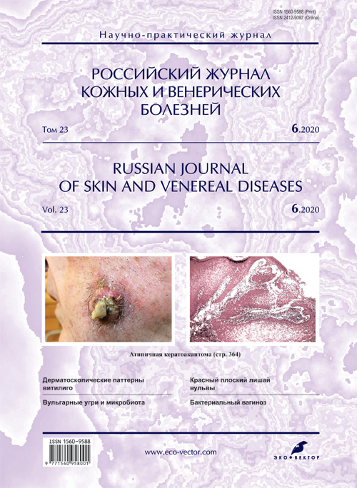
Пациент Ж., 73 года, болен в течение 7,5 мес. Заболевание началось с появления резко выступающего над окружающей кожей узелка розового цвета величиной с булавочную головку на коже левой щеки. Образование в течение 3 недель быстро увеличилось в размере. Субъективные ощущения варьировали в течении этого периода от зуда до резкой болезненности при касании, в последнее время отмечал умеренное кровление.
Локальный статус: на коже лица в центральной части левой щеки располагается солитарное образование в виде куполообразного крупного узла размером 2,1 × 2,3 см в диаметре, плотноэластической консистенции. В центральной части образования располагается центральный кратер, выступающий в виде гребня высотой 1,5 см, напоминающий кожный рог, окружённый светло-серыми роговыми массами. По периферии образования кожа местами синюшно-багрового цвета, множество жёстких волос. Валикообразный периферический край опухоли бледно-розового цвета, с телеангиэктазиями. Регионарные лимфатические узлы слева (подчелюстные) увеличены до размера лесного ореха (рис. 1).

Рис. 1. Больной Ж., 72 года. Атипичная кератоакантома кожи щеки в виде кожного рога

Рис. 2. Патоморфологическая картина кератоакантомы: акантолитические тяжи эпидермиса погружены в дерму на разную глубину, выражены явления псевдоэпителиоматозной гиперплазии. Окраска гематоксилином и эозином. Ув. 200
Результат гистологического исследования: патоморфологическая картина представлена пролиферирующими акантолитическими тяжами эпидермиса, погружёнными в дерму на различную глубину, с явлениями псевдоэпителиоматозной гиперплазии (рис. 2).
Клинический диагноз: атипичная кератоакантома в форме «кожного рога».
Пациента направили в онкологический центр, где опухоль была тотально иссечена. Наблюдение в сроки до 1 года, рецидива не выявлено.
Обсуждение
Несмотря на то, что большинство исследователей считают КА доброкачественной опухолью, есть убедительные доводы в пользу её трансформации в ПКР кожи, особенно в случаях атипичного характера течения процесса. Ряд авторов рассматривают КА как абортивное злокачественное новообразование или в качестве факультативного предрака [1, 2]. Следует учитывать тенденцию КА к быстрому росту, и как можно раньше выявлять атипичные формы КА не только с целью выбора рациональной терапевтической тактики, но и для профилактики злокачественной трансформации КА. В нашем клиническом наблюдении у пациента в возрасте 73 лет отмечался быстрый рост новообразования, который превышал сроки развития и регресса типичной КА (3 мес). Обращают на себя внимание и размеры опухоли, которые превышали 2 см, что говорит о гигантской форме новообразования. Кроме того, наличие центрального кратера, выступающего в виде гребня высотой 1,5 см, напоминающего кожный рог, окружённого светло-серыми роговыми массами, позволяет отнести этот вариант КА к атипичной КА по типу «кожного рога». По совокупности этих клинических признаков пациенту установлен окончательный диагноз и принято решение о гистологическом исследовании с целью исключения ПКР кожи.
Большинство исследователей предполагают радикальные методы лечения, так как считается, что персистирующие и рецидивирующие образования должны быть полностью удалены, особенно солитарные новообразования и опухоли на лице. Известны случаи рецидивирования КА после хирургического лечения, в том числе с элементами сосудистой и периневральной инвазии, что представляет необходимость динамического наблюдения, длительность которого определяется индивидуально с учётом возраста пациентов и доступности онкологической службы. Также имеет большое значение соблюдение профилактических мероприятий, направленных на предупреждение развития и рецидивирования КА. В случаях предрасположенности необходимо избегать прямых солнечных лучей и использовать фотопротекторы.
Заключение
Таким образом, в целях ранней диагностики и профилактики развития ПКР кожи диагноз КА должен устанавливать клиницист в сотрудничестве с патоморфологом и онкологом. Подход к лечению должен быть дифференцированным и базироваться на основании морфотипа опухоли.
Об авторах
Елена Сергеевна Снарская
ФГАОУ ВО «Первый Московский государственный медицинский университет им. И.М. Сеченова» Минздрава России (Сеченовский университет)
Автор, ответственный за переписку.
Email: snarskaya-dok@mail.ru
ORCID iD: 0000-0002-7968-7663
Scopus Author ID: 8714450500
доктор мед. наук, профессор, кафедра кожных и венерических болезней ФГАОУ ВО «Первый МГМУ им. И.М. Сеченова» Минздрава России
Россия, МоскваЛидия Мухамедовна Шнахова
ФГАОУ ВО «Первый МГМУ им. И.М. Сеченова» Минздрава России (Сеченовский Университет)
Email: Lika-slm@mail.ru
ORCID iD: 0000-0003-3000-0987
ассистент кафедры кожных и венерических болезней им. В.А. Рахманова
Россия, МоскваДарья Александровна Гомич
ФГАОУ ВО «Первый МГМУ им. И.М. Сеченова» Минздрава России (Сеченовский Университет)
Email: gomich28@icloud.com
ORCID iD: 0000-0002-4624-3046
Ординатор кафедры кожных и венерических болезней им. В.А. Рахманова
Россия, МоскваКсения Дмитриевна Васильева
ФГАОУ ВО «Первый МГМУ им. И.М. Сеченова» Минздрава России (Сеченовский Университет)
Email: kseniya07101988@mail.ru
ORCID iD: 0000-0001-8693-9622
Ординатор кафедры кожных и венерических болезней им. В.А. Рахманова
Россия, МоскваСписок литературы
- Галил-Оглы Г.А., Молочков В.А., Сергеев Ю.В., ред. Дерматоонкология. М.: Медицина для всех; 2005:241-300.
- Молочков В.А., Казанцева И.А., Кунцевич Ж.С., Бочкарева Е.В. Кератоакантома. М.: БИНОМ; 2006.
- Снарская Е.С., Молочков В.А. Базалиома. М.: Практическая медицина; 2018:48-69.
- Rook A., Whimster I. Keratoacanthoma – a thirty year retrospect. Br. J. Dermatol. 1979;100(1):41-7.
- Голдсмит Л.А., Кац С.И., Джилкрест Б.А., Паллер Э.С., Леффель Д.Дж., Вольф К. Дерматология Фицпатрика в клинической практике. Пер. с англ. М.: Издательство Панфилова; 2015. Т.2.
- Malcotti V., Kuroku K., Nahavama H., Doi K. Effects of double UVB-irradiations with different intervals on the dorsal skin of wistar-derived hypotrichotic WBN/ILA-Ht rats. Exp Toxicol Pathol. 2001;53(2-3):107-14.
- Олисова О.Ю., Андреева Е.В. Еще раз о проблеме гиперпигментации // Российский журнал кожных и венерических болезней. 2014;17(2):20-4.
- Олисова О.Ю., Владимирова Е.В., Бабушкин А.М. Кожа и солнце // Российский журнал кожных и венерических болезней. 2012;15(6):57-62.
- Corominas M., Sloan S.R., Leon J., Kamino H., Newcomb E.W., Pellicer A. Ras activation in human tumors and animal model systems. Environ Health Perspect. 1991;93:19-25. doi: 10.1289/ehp.919319
- Schwartz R.A., Klein E. Ultraviolet light-induced carcinogenesis. In: Holland J.F., Frei E III, eds. Cancer Medicine. Philadelphia: Lea&Febiger; 1982:109-19.
- Trowell H.E., Dyall-Smith M.L., Dyall-Smith D.J. Human papillomavirus associated with keratoacanthomas in Australian patient. Arch Dermatol. 1990;126(12):1654.
- Soler C., Chardonnet V., Euvrard S., Chignol M.C., Thivolet J. Evaluation of human papillomavirus type 5 on frozen sections of multiple lesions from transplant recipients with in situ hybridization and non-isotopic probes. Dermatology. 1992;184(4):248-53.
- Hsi E.D., Svoboda-Newman S.M., Stern R.A., Nickoloff B.J., Frank T.S. Detection of human papillomavirus DNA in keratoacanthomas by polymerase chain reaction. Am J Dermatopathol. 1997;19(1):10-5.
- Forslund O., De Angelis P.M., Beigi M., Schjolberg A.R., Clausen O.P. Identification of human papillomavirus in keratoacanthomas. J Cutan Pathol. 2003;30(7):423-9.
- Кладова А.Ю., Куевда Д.А., Кунцевич Ж.С., Прокофьев А.А., Багапш Л.С. К ассоциации кератоакантом с вирусом папилломы человека // Альманах клинической медицины. 2007;(15):187-91.
- Schwartz R.A. Keratoacanthoma. J Am Acad Dermatol. 1994;30(1):1-19.
- Hamilton S.A., Dickson W.A., O’Brein C.J. Keratoacanthoma developing in a split skin graft donor site. Br J Plast Surg. 1997;50(7):560-1.
- Олисова О.Ю., Додина М.И., Кушлинский Н.Е. Роль фактора роста сосудистого эндотелия в патогенезе розацеа и его медикаментозная коррекция // Клиническая дерматология и венерология. 2012;10(1):49-55.
- Lloyd K.M., Madsen D.K., Lin P.Y. Grzybowski’s eruptive keratoacanthoma. J Am Acad Dermatol. 1989;21(5, Pt1):1023-4.
- Okuyama R., Takahashi K., Ohi T., Tagami H. Keratoacanthoma developing in prurigo nodularis treated with cryotherapy. Dermatology. 1997;194(3):290-2.
- Chave T.A., Graham-Brown R.A. Keratoacanthoma developing in hypertrophic lichen planus. Br J Dermatol. 2003;148(3):592.
- Christofoletti Daldon P.E., Macado de Souza E., Cintra M.L. Hypertrophic lupus erythematosus:a clinicopathological study of 14 cases. J Cutan Pathol. 2003;30(7):443-8.
- Wiemers S., Stengel R., Schopf E., Laaff H. Subungual keratoacanthoma. Hautarzt. 1994;45(1):25-8.
- Akar A., Bulent Tastan H., Ozcan A., Erbil H., Riza Gur A. Multiple keratoacanthomas arising on skin lesions of pseudoxanthoma elasticum. J Eur Acad Dermatol. Venereol. 2002;16(5):533-4.
- Gheeraert P., Goens J., Schwartz R.A., Lambert W.C., Schroeder F., Debusscher L. Florid cutaneous papillomatosis, malignant acanthosis nigricans, and pulmonary squamous cell carcinoma. Int J Dermatol. 1991;30(3):193-7.
- Penmetcha M., Haighet A.S., Hopkinson J.M. Failure of PUVA in lichen myxoedematosus: acceleration of associated multiple keratoacanthomas with development of squamous carcinoma. Clin Exp Dermatol. 1987;12(3):220-3.
- Pellicano R., Fabrizi G., Cerimele D. Multiple keratoacanthomatas and junctional epidermlisis bullosa: a therapeutic conundrun. Arch Dermatol. 1990;126(3):305-6.
- Chaffai M., Houman M.H., Haouet S., Ben Osman A. Keratoacanthoma centrifugum marginatum. Ann Dermatol Venereol. 1994;121(10):731-3.
- Badell A., Marcoval J., Gallego I., Moreno A., Peyri J. Keratoacanthoma arising in hypertrophic lichen planus. Br J Dermatol. 2000;142(2):380-2.
- Kazakov D.V., Belousova I.E., Michaelis S., Palmedo G., Samtsov A.V., Kempf W. Unusual manifestation of specific cutaneous involvement by B-cell chronic lymphocytic leukemia: spontaneous regression with scar formation. Dermatology. 2003;207(1):111-5.
- Stoebner P.E., Fabre C., Delfour C., Joujoux J.M., Roger P., Dandurand Michel, Meunier L. Solitary subungual keratoacanthoma arising in an MSH2 germline mutation carrier: Confirmation of a relationship by immunohistochemical analysis. Dermatology. 2009;219(2):174-8.
- Le Boit P.E., Burg G., Weedon D., Sarasait A., eds. World Health Organization Classification of tumors. Pathology and genetics of skin tumors-IARC. Lyon: Press; 2006.
- Shcwartz R.A., Goldberg D.J., Mahmood F., DeJager R.L., Lambert W.C., Cohen P.J. The Muir-Torre syndrome: a disease of sebaceous and colonic neoplasms. Dermatologica. 1989;178(1):23-8.
- Хлебникова А.Н., Бочкарева Е.В., Гуревич Л.Е., Корсакова Н.А. Новые подходы к дифференциальной диагностике кератоакантомы и плоскоклеточного рака кожи // Российский журнал кожных и венерических болезней. 2009;11(5):4-10.
- Кунцевич Ж.С. Кератоакантома (дифференциальная диагностика с плоскоклеточным раком кожи, совершенствование методов лечения и профилактика озлокачествления): Автореф. дис. ….д-ра мед. наук. М.; 2007. Доступно на: https://www.elibrary.ru/item.asp?id=16163321; https://dlib.rsl.ru/01004054574
- Aramburu-Gonzalez J.A., Rodrigues-Justo M., Jimenez-Reyes J., Santonja C. A case of soft tissue mesenchymal chondrosarcoma metastatic to skin, clinically mimicking keratoacanthoma. Am J Dermatopathol. 1999;21(4):392-4.
- Saito M., Sasaki Y., Yamazaki N., Shimiru H. Self-involution of giant keratoacanthoma of the tip of the nose. Plast Reconstr Surg. 2003;111(4):1561-2.
- Narayan S., De Berker D., Oxey J. Ki-67 immunohistochemical staining distinguish subungualkeratoacanthoma from squamous cell carcinoma. In: Abstracts of the 20th World Congress of Dermatology. 1-5 July 2002, Paris, France. Ann Dermatol Venereol. 2002;129(Suppl 1, Pt 2):P0585.
- Asch P.H., Basset P., Roos M., Grosshans E., Bellocq J.P., Cribier B. Expression of stromelysin 3 in keratoacanthoma and squamous cell carcinoma. Am J Dermatopathol. 1999;21(2):146-50.
- Tran T.A., Ross J.S., Sheehan C.E., Carlson J.A. Comparison of oncostatin M expression in keratoacanthoma and squamous cell carcinoma. Mod Pathol. 2000;132(4):427-32.
- Рубина К.А., Сысоева В.Ю., Семина Е.В., Юрлова Е.И., Молочков В.А., Хлебникова А.Н., Седова Т.Г. Особенности экспрессии Т-кадгерина в кератиноцитах и сосудах эпителиальных опухолей кожи // Российский журнал кожных и венеричеcких болезней. 2013;15(1):9-15.
- Филоненко Е.В. Флюоресцентная диагностика и фотодинамическая терапия – обоснование применения и возможности в онкологии // Фотодинамическая терапия и фотодиагностика. 2014;(1):3-7.
- Кузнецов В.В. Использование фотодинамической терапии в отечественной онкологии (обзор литературы) // Исследования и практика в медицине. 2015;2(4):98-105.
- Сухова Т.Е., Молочков В.А., Романко Ю.С., Матвеева О.В., Решетников А.В. Лечение базальноклеточного рака кожи на современном этапе // Альманах клинической медицины. 2008;(18):14-21.
- Каплан М.А., Капинус В.Н., Романенко Ю.С., Ярославцева-Исаева Е.В. Фотодитазин – эффективный фотосенсибилизатор для фотодинамической терапии // Российский биотерапевтический журнал. 2004;3(2):50.
- Borovaya A., Olisova O., Ruzicka T., Sardy M. Does isotretinoin therapy of acne cure or cause depression? Inter J Dermatol. 2013;52(9):1040-52.
- Молочков В.А., Кунцевич Ж.С. К иммунотерапии и профилактике озлокачествления атипичных кератоакантом // Российский журнал кожных и венерических болезней. 2002;(3):4-8.
- Leonardi C., Zhu W.Y., Kinsey W.H., Penneys N.S. Seborrheic keratoses from the genital region may contain human papillomavirus DNA. Arch Dermatol. 1991;127(8):1203-6.
Дополнительные файлы
