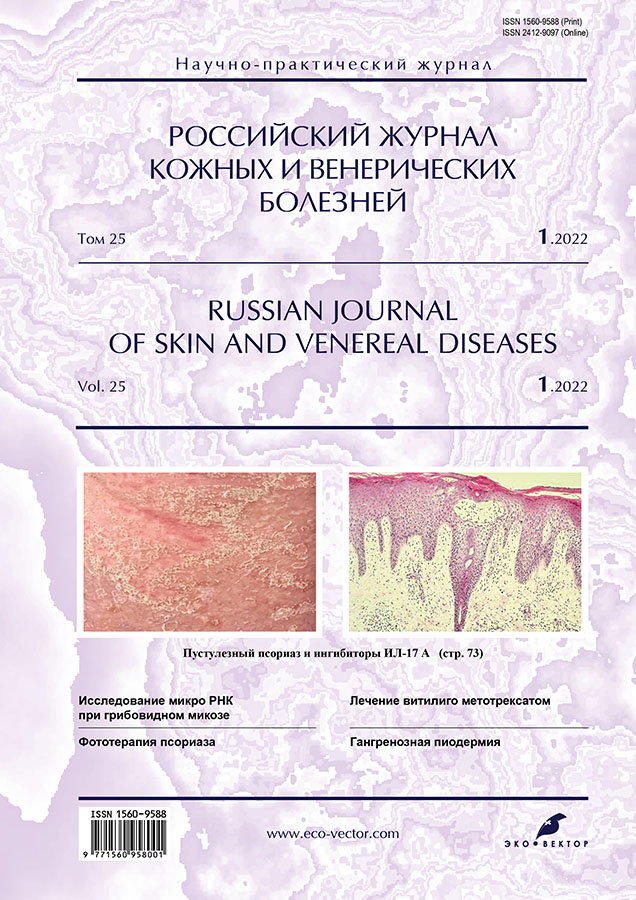
Том 25, № 1 (2022)
- Год: 2022
- Выпуск опубликован: 03.08.2022
- Статей: 9
- URL: https://rjsvd.com/1560-9588/issue/view/5375
- DOI: https://doi.org/10.17816/dv.251
Весь выпуск
ДЕРМАТООНКОЛОГИЯ
МикроРНК как диагностический маркер при Т-клеточных лимфомах кожи
Аннотация
Обоснование. В последние годы благодаря развитию методов молекулярно-генетического анализа одним из перспективных маркеров для диагностики многих заболеваний человека становится микроРНК.
Цель ― изучение микроРНК в плазме крови для ранней диагностики грибовидного микоза.
Материал и методы. В исследование были включены 30 пациентов с гистологически подтверждённым диагнозом Т-клеточной лимфомы кожи, у 25 был диагностирован грибовидный микоз, у 5 ― синдром Сезари. Группу контроля составили 10 пациентов с доброкачественными лимфопролиферативными дерматозами. Пациентам проводилось определение микроРНК 223, 16, 326, 663, 423, 711 в плазме крови. Проведено также определение микроРНК в плазме у пациентов с Т-клеточными лимфомами кожи на ранних и поздних стадиях.
Результаты. Выявлена статистически значимая разница микроРНК 223, 16, 326, 711 в плазме крови у пациентов с грибовидным микозом в сравнении с пациентами с доброкачественными лимфопролиферативными дерматозами, микроРНК 663 у пациентов с Т-клеточными лимфомами кожи на ранней и поздней стадиях, а также микроРНК 223, 711 у пациентов с грибовидным микозом на ранней стадии в сравнении с пациентами с доброкачественными лимфопролиферативными дерматозами.
Заключение. Определение микроРНК 223, 16, 326, 711 в плазме крови может быть использовано для ранней диагностики Т-клеточной лимфомы кожи.
 5-16
5-16


ДЕРМАТОЛОГИЯ
Клинико-патогенетическое обоснование применения метотрексата в лечении прогрессирующего несегментарного витилиго
Аннотация
Обоснование. Витилиго является актуальной проблемой как для пациентов, так и для научного сообщества дерматологов. Проводится множество исследований, направленных на поиск новых методов терапии данного заболевания, однако схем лечения, обеспечивающих репигментацию очагов и стабилизацию процесса, нет. В связи с этим актуален поиск средств, оказывающих патогенетическое действие.
Цель ― клинико-лабораторная оценка эффективности применения метотрексата при витилиго.
Материал и методы. Представлены предварительные результаты исследования эффективности метотрексата при лечении витилиго. Клинические эффекты оценивались с использованием индекса VES (оценка степени витилиго). Определены также динамика иммунологических показателей и влияние витилиго на качество жизни.
В исследование было включено 77 пациентов, страдающих несегментарным витилиго. Все исследуемые были разделены на 2 группы. Пациенты 1-й группы (39 больных) получали метотрексат в сочетании с фототерапией, пациентам 2-й группы (38 больных) проведён курс фототерапии с использованием ультрафиолетовых лучей типа В с длинной волны 311 нм (УФБ-311 нм). Определялась площадь депигментации, а после лечения ― репигментации относительно площади поверхности тела. Анализировалась динамика иммунного статуса на фоне проводимой терапии. Оценивалась корреляция распространённости процесса с качеством жизни. Продолжительность исследования составила 4 мес.
Результаты. Статистически значимых различий до лечения по клиническим и лабораторным показателям среди групп не выявлено. Пациенты 1-й группы (комбинированная терапия метотрексатом в сочетании со средневолновой узкополосной терапией УФБ-311 нм) продемонстрировали не только более активную репигментацию очагов витилиго, чем исследуемые 2-й группы, но и наиболее активную положительную динамику дерматологического индекса качества жизни, коррелирующую с распространённостью процесса. Анализ динамики показателей иммунного статуса позволил сделать вывод о наилучшей тенденции к нормализации уровня цитокинов среди пациентов 1-й группы.
Заключение. Проведённое исследование продемонстрировало клиническую и патогенетическую эффективность метотрексата. Малые дозы препарата хорошо переносятся, что позволяет длительно воздействовать на патогенетические механизмы, стабилизируя кожный процесс.
 17-27
17-27


Фототерапия псориаза
Аннотация
Псориаз ― хроническое генетически детерминированное заболевание мультифакториальной природы, связанное с иммуноопосредованным воспалением и характеризующееся рецидивирующим течением с частым ассоциативным поражением других органов и систем.
Согласно общемировым рекомендациям, в настоящее время, несмотря на наличие широкого выбора новейших таргетных генно-инженерных биологических препаратов, фототерапия продолжает занимать важную нишу в лечении среднетяжёлого и тяжёлого псориаза благодаря своему патогенетически обоснованному целенаправленному действию, безопасности и низкой стоимости процедур.
В обзоре представлены подробные данные о механизме действия, эффективности фототерапии, а также о потенциальных биомаркерах псориаза (кальпротектин, липокалин 2, резистин), с помощью которых с высокой долей вероятности будет дана точная оценка результативности проводимого лечения и при необходимости его своевременная коррекция, что позволит быстрее достигать желаемого эффекта и, как следствие, повышать качество жизни пациентов.
 29-39
29-39


Сочетание актинической, гипертрофической и типичной форм красного плоского лишая у одного пациента
Аннотация
Красный плоский лишай ― хронически протекающий дерматоз мультифакториальной природы, для которого характерно появление плоских полигональных зудящих папул на коже и слизистых оболочках. Дерматоз часто ассоциирован с сахарным диабетом и заболеваниями желудочно-кишечного тракта, крайне редко ― с онкологическими заболеваниями. Для лечения рекомендуются антималярийные препараты, обладающие фотозащитным, противовоспалительным, слабым иммунодепрессивным эффектом.
Актиническая и гипертрофическая формы красного плоского лишая относятся к атипичным формам заболевания. Актинический или тропический красный плоский лишай в Российской Федерации встречается очень редко, в основном в странах Среднего и Ближнего Востока, Средней Азии, Африки. Актиническая форма красного плоского лишая характеризуется локализацией на открытых участках кожи (лицо, шея). Для гипертрофической формы красного плоского лишая характерны папулы больших размеров с бугристой поверхностью, которые совсем не похожи на таковые при типичной форме. Редкая встречаемость дерматоза и необычная локализация приводят к затруднению постановки диагноза.
В статье приводится описание клинического случая сочетания актинической, гипертрофической и типичной форм красного плоского лишая у пациента, родившегося на Кавказе, но длительное время проживающего в России. Кожный процесс у пациента носил распространённый характер, был локализован по всему кожному покрову, включая лицо, шею, слизистые оболочки полости рта; свободными от высыпаний оставались лишь кожа ладоней и подошв. Сыпь была представлена плоскими папулами полигональной формы, синюшно-розового цвета, размером с чечевицу, и белесоватой сеткой Уикхема на поверхности.
Локализация на открытых участках кожи, сочетание с гипертрофическими папулами, работа на открытом воздухе, сильный зуд с экскориациями привели к неправильной постановке диагноза, а проводимая терапия не давала эффекта. Для уточнения диагноза было проведено гистологическое исследование, выявившее выраженный гиперкератоз, неравномерный гранулёз, массивный папилломатоз в сосочковом и подсосочковом слоях дермы (полосовидный умеренный инфильтрат из лимфоидных элементов, гистиоцитов, незначительный отёк, расширение сосудов). Адекватно подобранная медикаментозная терапия (дексаметазон внутримышечно; хлоропирамин внутримышечно; противомалярийное средство; никотиновая кислота; наружно дерматоловая мазь), а также сеансы иглорефлексотерапии вторым тормозным методом привели к улучшению состояния.
По завершении лечения пациенту рекомендованы фотозащитные наружные средства на открытые участки кожи и повторный курс акупунктуры.
 41-47
41-47


Декальвирующий фолликулит: клинико-морфологическая характеристика (обзор литературы)
Аннотация
Декальвирующий фолликулит ― редкое заболевание из группы первичных рубцовых алопеций, на долю которого приходится 11% всех алопеций данной группы. Дерматоз впервые был описан французским врачом-дерматологом Charles-Eugène Quinquaud в 1888 и 1889 гг. В последние десятилетия происходит увеличение количества публикаций, посвящённых описанию этиопатогенеза, клинических и гистологических характеристик, а также подходов к терапии декальвирующего фолликулита.
В статье представлены результаты анализа данных по базам Scopus, Web of Science, MedLine, the Cochrane Library, EMBASE, Global Health, CyberLeninka, РИНЦ.
Этиопатогенез заболевания до настоящего времени неизвестен. Ранее обсуждались роль себореи и колонизация кожи Staphylococcus aureus, а также нарушение локального иммунного ответа и наличие генетической предрасположенности. В настоящее время считается, что при декальвирующем фолликулите происходит значимое и персистирующее нарушение кожного барьера, которое предрасполагает к субэпидермальной инвазии оппортунистических микроорганизмов, в том числе Staphylococcus aureus.
Клинические, дерматоскопические (трихоскопические) и гистологические характеристики дерматоза уточняются. Характерными клиническими чертами его являются упорное прогрессирующее течение, формирование очагов алопеции с насыщенно-красным краем, пустулами и корками по периферии очагов алопеции, политрихией и образованием плотного рубца, возвышающегося над окружающей кожей. Дерматоскопические характеристики находятся в зависимости от степени воспалительного процесса. К специфическим трихоскопическим признакам заболевания относят фолликулярные пустулы, жёлтое тубулярное шелушение, жёлтые корки, перифолликулярную эритему, перифолликулярные геморрагии и тонкие извитые сосуды. В зависимости от количества данных признаков определяется степень воспаления. Гистологические черты заболевания включают массивный перифолликулярный инфильтрат, формирование щелей между эпителием фолликулов и окружающей стромой, а на финальных стадиях процесса ― фиброзные тракты, диффузный фиброз в дерме.
Препаратами выбора для лечения декальвирующего фолликулита являются антибактериальные препараты, также возможна терапия курсами топических кортикостероидов, антисептических растворов.
Предполагаем, что систематизация сведений об этиопатогенезе и подходах к диагностике и лечению декальвирующего фолликулита послужит улучшению диагностики среди других первичных рубцовых алопеций и выбору тактики лечения дерматоза.
 49-59
49-59


Гангренозная пиодермия: опыт обследования и лечения
Аннотация
Обоснование. Гангренозная пиодермия ― редкое воспалительное заболевание кожи, которое в настоящее время относится к группе нейтрофильных дерматозов.
Цель ― разработка эпидемиологических, клинических и лабораторных характеристик пациентов с гангренозной пиодермией, а также протокола лечения этого заболевания.
Материал и методы. Представлены результаты одноцентрового когортного проспективного исследования, которое проводилось на базе клиники кожных и венерических болезней имени В.А. Рахманова Первого МГМУ имени И.М. Сеченова в период с января 2019 года по ноябрь 2021 года, включая всех пациентов с подтверждённым диагнозом гангренозной пиодермии.
Результаты. В результате исследования выявлено, что 16 (53%) пациентов из 30 были женщинами, средний возраст на момент постановки диагноза составлял 59±16,3 лет. Наиболее частой локализацией высыпаний являлась голень (20 пациентов; 67%), редко (по одному случаю) отмечалось поражение кожи лица, половых органов, молочных желёз. У 14 (47%) пациентов выявлялись одномоментно две и более язв (максимально 9 язв). Феномен патергии был положительный у 23 (77%) пациентов из 30. Язвенная форма гангренозной пиодермии была у 25 (83%), у одного пациента выявлена внекожная форма заболевания с поражением лёгких. Наиболее частое ассоциированное заболевание ― ревматоидный артрит ― отмечен у 6 (20%) пациентов; редкими ассоциированными заболеваниями (по одному наблюдению) были также гепатит С (у 2), синдром множественной эндокринной неоплазии 1-го типа, аутоиммунный гепатит, неходжскинская лимфома. При гистологическом исследовании нейтрофильная инфильтрация дермы выявлялась в 100% случаев, а лейкоцитокластический васкулит ― в 53% (у 16). Полное рубцевание очагов на фоне проведённого лечения наблюдалось у 22 (73%) пациентов. За период наблюдения рецидивы заболевания имели место у 12 (40%) пациентов, в том числе два летальных исхода.
Заключение. Представлена одна из крупнейших серий случаев гангренозной пиодермии на сегодняшний день. В процессе обследования больных выявлено, что нейтрофильная инфильтрация дермы является характерным признаком заболевания. Обнаружены редкие ассоциированные заболевания (синдром множественной эндокринной неоплазии 1-го типа, аутоиммунный гепатит, гепатит С, неходжскинская лимфома). Достаточно большой процент рецидивов свидетельствует о необходимости дальнейших исследований с целью разработки дополнительного метода обследования для современной диагностики и обоснования своевременного назначения препаратов таргетной терапии.
 61-72
61-72


Эффективность ингибитора IL-17А при генерализованном пустулёзном псориазе: клинический случай
Аннотация
Представлен первый клинический случай эффективного применения отечественного биологического препарата ― ингибитора IL-17А нетакимаба ― у больного упорно прогрессирующим, торпидным к терапии генерализованным пустулёзным псориазом Цумбуша. Этот редкий системный дерматоз относится к тяжёлым формам псориаза, угрожающим жизни пациента и требующим проведения интенсивной терапии уже с первых часов проявления заболевания. Развитию пустулёзного псориаза может способствовать длительная терапия системными глюкокортикостероидами, цитостатиками, пероральными контрацептивами, а также длительное использование раздражающих наружных средств. В некоторых случаях заболевание связывают с высокими эмоциональными нагрузками, стрессами. При тяжёлом генерализованном пустулёзном псориазе эффективными оказываются биологические препараты, циклоспорин, метотрексат, ацитретин.
В статье представлены сводные данные об эффективности препаратов первой и второй линии в лечении пустулёзного псориаза. По данным литературы, пациенты с тяжёлым пустулёзным псориазом, торпидные к стандартной терапии, демонстрируют положительный ответ на лечение биологическими препаратами в большинстве случаев. Анти-TNF-α являются наиболее доступными биологическими препаратами для лечения пустулёзного псориаза, а анти-IL-12/23 и анти-IL-17A могут рассматриваться в качестве первой или второй линии терапии при умеренно тяжёлом и рефрактерном пустулёзном псориазе.
Выбор эффективной терапии для лечения пустулёзного псориаза является актуальной проблемой современной дерматологии. Представленный клинический случай демонстрирует эффективность применения ингибитора IL-17А и выраженный положительный терапевтический эффект уже в течение одних суток в лечении резистентного к ранее проводимой терапии генерализованного пустулёзного псориаза, что позволяет рассчитывать на перспективу дальнейшего успешного применения этого таргетного препарата.
 73-83
73-83


ХРОНИКА
Хроника Московского общества дерматовенерологов и косметологов имени А.И. Поспелова (МОДВ основано 4 октября 1891 г.) Бюллетень заседания МОДВ № 1146
Аннотация
15 февраля 2022 года состоялось 1146-е заседание Московского общества дерматовенерологов и косметологов имени А.И. Поспелова.
Встреча проходила в удалённом формате. Всего присутствовало 108 участников. Принят в члены МОДВ один кандидат.
Представлены три клинических наблюдения. Первое из них ― случай гигантоклеточной кольцевидной гранулёмы с обширным поражением кожи. Кольцевидная гранулёма ― доброкачественный дерматоз неясной этиологии, склонный к самопроизвольному разрешению; возникает как реакция на ультрафиолетовое излучение на фоне лимфопролиферативных или аутоиммунных заболеваний, саркоидоза, хронических заболеваний лёгких, рака простаты. В представленном случае клинический диагноз подтверждён гистологическим исследованием.
Второе выступление было посвящено ливедоидной васкулопатии со склонностью к тромботическим осложнениям. Это относительно редкое заболевание, которое исключено из группы васкулитов из-за одной отличительной особенности ― выраженной склонности к тромбообразованию при отсутствии указаний на первичный характер воспаления. В терапии кроме медикаментозных средств обязательным является назначение витаминов.
Третье наблюдение касалось карциномы Меркеля ― одной из относительно редких, но крайне агрессивных форм рака кожи с эпителиальной и нейроэндокринной дифференцировкой, которая по агрессивности течения и склонности к метастазированию превосходит меланому. Из-за редкой встречаемости опухоли часты ошибки диагностики. Диагностика требует гистологического и иммуногистохимического исследования. Любая «воспалившаяся атерома» или синовиальная киста, рецидивирующая после хирургического лечения, должна настораживать.
Научные доклады были посвящены биологической терапии псориаза ― хронического системного иммуноассоциированного заболевания мультифакториальной природы с наследственной предрасположенностью, характеризуемого образованием воспалительных папул и бляшек, часто с поражением суставов (артропатия), ускоренной пролиферацией эпидермоцитов и нарушением их дифференцировки, дисбалансом между провоспалительными и противовоспалительными цитокинами и хемокинами. Второй научный доклад содержал информацию о современных подходах к лечению акантолитической пузырчатки.
 85-91
85-91


ФОТОГАЛЕРЕЯ
Фотогалерея. Аногенитальные (венерические) бородавки
Аннотация
Аногенитальные бородавки обусловлены вирусом папилломы человека (наиболее часто 6-м и 11-м, гигантские кондиломы ― 16-м и 18-м типами) и представляют собой экзо- и эндофитные разрастания на коже и слизистых оболочках половых органов и перианальной области. Общепринятой классификации нет, однако, исходя из клинических проявлений, выделяют остроконечные кондиломы, папулёзные, пятнистые, гиперкератотические, гигантские кондиломы Бушке–Левенштейна. Аногенитальные бородавки могут быть следствием инфицирования при сексуальных контактах, на что и указывает синоним «венерические бородавки». Аногенитальные бородавки представляют собой отдельную нозологию (шифр по МКБ-10 А63.0), но могут быть и частью симптомокомплекса при иммунодефицитных состояниях (в частности, при синдроме приобретённого иммунодефицита) и неоплазиях (плоскоклеточном раке и эритроплазии Кейра).
Предлагаем публикацию фотогалереи по данной проблеме.
 93-96
93-96


