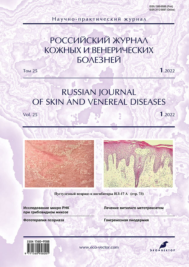
Фотогалерея. Аногенитальные (венерические) бородавки
- Авторы: Дубенский В.В.1, Дубенский В.В.1
-
Учреждения:
- Тверской государственный медицинский университет
- Выпуск: Том 25, № 1 (2022)
- Страницы: 93-96
- Раздел: ФОТОГАЛЕРЕЯ
- Статья получена: 30.03.2022
- Статья одобрена: 03.05.2022
- Статья опубликована: 03.08.2022
- URL: https://rjsvd.com/1560-9588/article/view/105693
- DOI: https://doi.org/10.17816/dv105693
- ID: 105693
Цитировать
Полный текст
Аннотация
Аногенитальные бородавки обусловлены вирусом папилломы человека (наиболее часто 6-м и 11-м, гигантские кондиломы ― 16-м и 18-м типами) и представляют собой экзо- и эндофитные разрастания на коже и слизистых оболочках половых органов и перианальной области. Общепринятой классификации нет, однако, исходя из клинических проявлений, выделяют остроконечные кондиломы, папулёзные, пятнистые, гиперкератотические, гигантские кондиломы Бушке–Левенштейна. Аногенитальные бородавки могут быть следствием инфицирования при сексуальных контактах, на что и указывает синоним «венерические бородавки». Аногенитальные бородавки представляют собой отдельную нозологию (шифр по МКБ-10 А63.0), но могут быть и частью симптомокомплекса при иммунодефицитных состояниях (в частности, при синдроме приобретённого иммунодефицита) и неоплазиях (плоскоклеточном раке и эритроплазии Кейра).
Предлагаем публикацию фотогалереи по данной проблеме.
Ключевые слова
Полный текст
Рис. 1. Больной С., 24 года. Диагноз: «Аногенитальные бородавки головки полового члена и внутреннего листка крайней плоти». Имеются экзофитные разрастания в виде остроконечных (а) и папулёзных кондилом (b) розового цвета, с чёткими границами; отдельные образования на ножке, склонные к росту, залегают на фоне неизменённой кожи.
Fig. 1. Patient C., 24 years old. Diagnosis: “Anogenital warts of the glans and inner foreskin”. There are exophytic growths in the form of acute (a) and papular condylomas (b) of pink color, with clear boundaries; separate formations on the pedicle, prone to growth, are located on the background of unchanged skin.
Рис. 2. Больной В., 32 года. Диагноз: «Аногенитальные бородавки внутреннего листка крайней плоти и уздечки полового члена». Разрастания в виде плоских папул с общим широким основанием напоминают «цветную капусту», имеют чёткие границы, залегают на фоне неизменённой кожи.
Fig. 2. Patient V., 32 years old. Diagnosis: “Anogenital warts of the inner foreskin and frenulum of the penis”. The growths were in the form of flat papules with a wide base, resembling “cauliflower”. They have clear borders, occur against the background of unchanged skin.
Рис. 3. Больная А., 21 год. Диагноз: «Аногенитальные бородавки наружных гениталий». Экзофитные разрастания в виде папулёзных и остроконечных кондилом располагаются в области больших и малых половых губ, имеют чёткие границы и сосочковидную поверхность.
Fig. 3. Patient A., 21 years old. Diagnosis: “Anogenital warts of the external genitalia”. Exophytic outgrowths in the form of papular and acute condylomas, located in the area of large and small labia, have clear borders and papilla-shaped surface.
Рис. 4. Больная К., 24 года. Диагноз: «Аногенитальные бородавки шейки матки». Папулёзные кондиломы на шейке матки с распространением в цервикальный канал, белесоватого цвета (цвет изменился после диагностической пробы с 3% раствором уксусной кислоты).
Fig. 4. Patient K., 24 years old. Diagnosis: “Anogenital warts of the cervix”. Papular condylomas on the cervix with spreading to the cervical canal, whitish color (color changed after diagnostic test with 3% acetic acid solution).
Рис. 5. Та же больная. Аногенитальные бородавки в преддверии влагалища. Остроконечные кондиломы розового цвета с чёткими границами на фоне видимо неизменённой слизистой оболочки.
Fig. 5. The same patient. Anogenital warts in the vaginal fornix. Pink acute condylomas with clear borders on the background of visibly unchanged mucosa.
Рис. 6. Больная Е., 28 лет. Диагноз: «Аногенитальные бородавки губок уретры. Остроконечные кондиломы красного цвета расположены на фоне неизменённой слизистой оболочки наружного отверстия уретры.
Fig. 6. Patient E., 28 years old. Diagnosis: “Anogenital warts of the urethral labia”. Red acute condylomas, located against the background of the unchanged mucosa of the external orifice of the urethra.
Рис. 7. Больной Т., 24 года. Диагноз: «Аногенитальные бородавки наружного отверстия мочеиспускательного канала». Экзофитные разрастания остроконечных кондилом в области губок уретры (а); сопутствующая венозная аневризма головки полового члена (b).
Fig. 7. Patient T., 24 years old. Diagnosis: “Anogenital warts of the external opening of the urethra”. Exophytic growths of acute condylomas in the region of the urethral labia (a); associated venous aneurysm of the penile head (b).
Рис. 8. Больной М., 23 года. Диагноз: «Аногенитальные бородавки (гигантские кондиломы Бушке–Левенштейна) кожи лобка и полового члена». Экзофитные опухолевидные разрастания розово-красного и буроватого цвета с отдельными сосочковидными образованиями.
Fig. 8. Patient M., 23 years old. Diagnosis: “Anogenital warts (Buschke–Lowenstein tumor) of the pubic and penile skin”. Exophytic tumor-like outgrowths of pink-red and brownish color, with separate papillae.
Рис. 9. Больной Ж., 27 лет. Диагноз: «Аногенитальные бородавки губок уретры». Сопутствующий диагноз: «Эритроплазия Кейра». Остроконечные кондиломы красного цвета с чёткими границами в области губок уретры (а) залегают на фоне неизменённой кожи и эритематозно-папулёзных участков эритроплазии (b).
Fig. 9. Patient J., 27 years old. Diagnosis: “Anogenital warts of the urethral labia”. Concomitant diagnosis: “Erythroplasia of Queyrat”. There are red acute condylomas with clear borders in the area of the urethral labia (a) they lie against the background of unchanged skin and erythematous-papular areas of erythroplasia (b).
Рис. 10. Больная Т., 36 лет. Диагноз: «Аногенитальные бородавки (гиперкератотические и гигантские кондиломы Бушке–Левенштейна) наружных гениталий, перианальной области и паховых складок». Сопутствующий диагноз: «Плоскоклеточный рак в области больших половых губ и нижней спайки». Имеются отдельные буровато-коричневого цвета гиперкератотические образования на коже паховых складок и больших половых губ (а). На малых и больших половых губах и в перианальной области располагаются гигантские кондиломы Бушке–Левенштейна (b) красно-синюшного цвета, с дольчатым делением на поверхности. В области нижней спайки имеется болезненный плотный инфильтрат красно-чёрного цвета, размером до 4 см в диаметре (с). Паховые лимфатические узлы увеличены с обеих сторон размером до «боба», болезненные при пальпации.
Fig. 10. Patient T., 36 years old. Diagnosis: “Anogenital warts (hyperkeratotic and Buschke–Lowenstein tumors) of the external genitalia, perianal area and inguinal folds”. Concomitant diagnosis: “Squamous cell carcinoma in the area of the labia majora and the lower commissure”. There were isolated brownish hyperkeratotic lesions on the skin of the inguinal folds and labia majora (a). There are Buschke–Lowenstein tumors on the labia majora and perianal region (b), reddish-brown, with lobular separation on the surface. There is a painful red-black dense infiltrate, up to 4 cm in diameter, in the area of the lower adhesion (c). The inguinal lymph nodes are enlarged on both sides, up to the size of a “bean”, painful on palpation.
Рис. 11. Больной Р., 2 года. Диагноз: «Аногенитальные бородавки перианальной области». Остроконечные кондиломы расположены симметрично на «целующихся» поверхностях, мацерированы, розово-красного цвета.
Fig. 11. Patient R., 2 years old. Diagnosis: “Anogenital perianal warts”. The acute condylomas are symmetrically located on the “kissing” surfaces, macerated, pink-red in color.
Об авторах
Валерий Викторович Дубенский
Тверской государственный медицинский университет
Email: valerydubensky@yandex.ru
ORCID iD: 0000-0003-2674-1096
SPIN-код: 3577-7335
Россия, Тверь
Владислав Валерьевич Дубенский
Тверской государственный медицинский университет
Автор, ответственный за переписку.
Email: dubensky.vladislav@yandex.ru
ORCID iD: 0000-0002-5583-928X
SPIN-код: 6044-8507
Россия, Тверь
Список литературы
Дополнительные файлы


